

In vitro and in vivo anti-tumor activities of anti-EGFR single-chain variable fragment fused with recombinant gelonin toxin
- PMID: 22392077
- PMCID: PMC11824807
- DOI: 10.1007/s00432-012-1181-7
In vitro and in vivo anti-tumor activities of anti-EGFR single-chain variable fragment fused with recombinant gelonin toxin
Abstract
Purpose: Epidermal growth factor receptor (EGFR) plays an important role in the growth and metastasis of many solid tumors. Strategies that target EGFR hold promising therapeutic potential for the treatment for non-small cell lung cancer (NSCLC), as EGFR is normally overexpressed in these tumors. This study was designed to determine whether an anti-EGFR immunotoxin has anti-tumor activity against NSCLC, and if so, to further investigate the possible mechanisms of cytotoxicity.
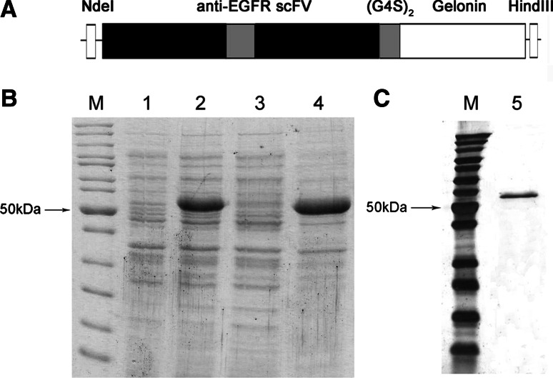
Methods: A fusion protein of anti-EGFR single-chain variable fragment (anti-EGFR scFv) and the plant toxin gelonin (rGel) was constructed, expressed in bacteria, and purified to homogeneity. Cytotoxicity of anti-EGFR scFv/rGel (E/rG) immunotoxin was assessed on A549, HCC827, and H1975 cells (EGFR-overexpressing NSCLC-derived cell lines) and A549 xenografts in nude mice.
Results: Cytotoxicity experiments using E/rG on A549, HCC827, and H1975 cells demonstrated that E/rG can specifically inhibit proliferation of these cells, whereas it did not affect the proliferation of Raji cells that do not express EGFR. Treatment for A549 xenografts in nude mice with E/rG resulted in significant suppression of tumor growth compared to controls. Immunofluorescence in frozen tissue sections confirmed that E/rG could specifically bind to tumor tissues in nude mice bearing A549 tumor xenografts, while rGel alone showed no binding activity. Furthermore, E/rG inhibited the growth of A549 cells by cytotoxic effects that blocked tumor proliferation, and the immunotoxin-induced cell death may be mediated by autophagy.
Conclusions: These results showed that E/rG might have significant potential as a novel clinical therapeutic agent against human NSCLC.
Conflict of interest statement
The authors declare that they have no conflict of interest.
Figures


References
-
- Allen TM (2002) Ligand-targeted therapeutics in anticancer therapy. Nat Rev Cancer 2(10):750–763 - PubMed
-
- Altekruse SF, Kosary CL, Krapcho M, Neyman N, Aminou R, Waldron W, Ruhl J, Howlader N, Tatalovich Z, Cho H, Mariotto A, Eisner MP, Lewis DR, Cronin K, Chen HS, Feuer EJ, Stinchcomb DG, Edwards BK (eds) (2009) SEER Cancer statistics review, 1975–2007. National Cancer Institute, Bethesda
-
- Baluna R, Sausville EA, Stone MJ, StetlerStevenson MA, Uhr JW, Vitetta ES (1996) Decreases in levels of serum fibronectin predict the severity of vascular leak syndrome in patients treated with ricin A chain-containing immunotoxins. Clin Cancer Res 2(10):1705–1712 - PubMed
-
- Borghaei H, Langer CJ, Millenson M, Ruth KJ, Litwin S, Tuttle H, Seldomridge JS, Rovito M, Mintzer D, Cohen R, Treat J (2008) Phase II study of paclitaxel, carboplatin, and cetuximab as first line treatment, for patients with advanced non-small cell lung cancer (NSCLC) results of OPN-017. J Thorac Oncol 3(11):1286–1292 - PubMed
-
- Brekke OH, Sandlie I (2003) Therapeutic antibodies for human diseases at the dawn of the twenty-first century. Nat Rev Drug Discov 2(1):52–62 - PubMed
Publication types
MeSH terms
Substances
LinkOut - more resources
Full Text Sources
Other Literature Sources
Medical
Research Materials
Miscellaneous
