Comparison of cardiac MRI tissue tracking and myocardial tagging for assessment of regional ventricular strain
- PMID: 22392105
- PMCID: PMC6941669
- DOI: 10.1007/s10554-012-0035-3
Comparison of cardiac MRI tissue tracking and myocardial tagging for assessment of regional ventricular strain
Abstract
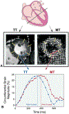
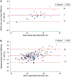
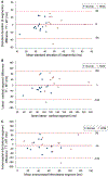
This study sought to compare regional measures of ventricular strain by tissue tracking (TT) to those derived from myocardial tagging (MT) within cardiac MR (CMR), in normal subjects and patients with hypertrophic cardiomyopathy. CMR images from 13 normal subjects and 11 subjects with hypertrophic cardiomyopathy were retrospectively analyzed. For each subject, equivalent mid-papillary level short-axis cine steady-state free precession and MT slices from the same examination were evaluated. The time to peak circumferential strain and magnitude of the peak strain were calculated for 6 matched left ventricular segments. Data from 24 slices (n = 144 segments) were compared. The mean difference between techniques in magnitude of peak strain and time to peak strain was 1 ± 9% and 1 ± 58 ms, respectively. The mean difference in the standard deviation of time to peak strain within a slice was 0 ± 19 ms (mean cardiac cycle duration 1,013 ± 204 ms). Bland-Altman analysis showed closer agreement in time to peak strain than peak strain magnitude. Measurements of segmental time to peak strain by TT and MT were in close agreement; agreement for the magnitude of peak segmental strain was more modest. The TT approach does not add to CMR examination time and may be a useful tool for the assessment of ventricular synchrony.
Figures


Similar articles
-
Comparison of left ventricular strains and torsion derived from feature tracking and DENSE CMR.J Cardiovasc Magn Reson. 2018 Sep 13;20(1):63. doi: 10.1186/s12968-018-0485-4. J Cardiovasc Magn Reson. 2018. PMID: 30208894 Free PMC article.
-
Feature tracking compared with tissue tagging measurements of segmental strain by cardiovascular magnetic resonance.J Cardiovasc Magn Reson. 2014 Jan 22;16(1):10. doi: 10.1186/1532-429X-16-10. J Cardiovasc Magn Reson. 2014. PMID: 24450803 Free PMC article.
-
Global and regional left ventricular myocardial deformation measures by magnetic resonance feature tracking in healthy volunteers: comparison with tagging and relevance of gender.J Cardiovasc Magn Reson. 2013 Jan 18;15(1):8. doi: 10.1186/1532-429X-15-8. J Cardiovasc Magn Reson. 2013. PMID: 23331550 Free PMC article.
-
Simultaneous strain-volume analysis by three-dimensional echocardiography: validation in normal subjects with tagging cardiac magnetic resonance.J Cardiovasc Med (Hagerstown). 2017 Apr;18(4):223-229. doi: 10.2459/JCM.0000000000000336. J Cardiovasc Med (Hagerstown). 2017. PMID: 26702593
-
Strain analysis in CRT candidates using the novel segment length in cine (SLICE) post-processing technique on standard CMR cine images.Eur Radiol. 2017 Dec;27(12):5158-5168. doi: 10.1007/s00330-017-4890-0. Epub 2017 Jun 27. Eur Radiol. 2017. PMID: 28656465 Free PMC article.
Cited by
-
Comparison of left ventricular strains and torsion derived from feature tracking and DENSE CMR.J Cardiovasc Magn Reson. 2018 Sep 13;20(1):63. doi: 10.1186/s12968-018-0485-4. J Cardiovasc Magn Reson. 2018. PMID: 30208894 Free PMC article.
-
Age- and sex-specific reference values of biventricular strain and strain rate derived from a large cohort of healthy Chinese adults: a cardiovascular magnetic resonance feature tracking study.J Cardiovasc Magn Reson. 2022 Nov 21;24(1):63. doi: 10.1186/s12968-022-00881-1. J Cardiovasc Magn Reson. 2022. PMID: 36404299 Free PMC article.
-
Cardiovascular imaging 2012 in the International Journal of Cardiovascular Imaging.Int J Cardiovasc Imaging. 2013 Apr;29(4):725-36. doi: 10.1007/s10554-013-0216-8. Int J Cardiovasc Imaging. 2013. PMID: 23589003 Review. No abstract available.
-
Feature tracking compared with tissue tagging measurements of segmental strain by cardiovascular magnetic resonance.J Cardiovasc Magn Reson. 2014 Jan 22;16(1):10. doi: 10.1186/1532-429X-16-10. J Cardiovasc Magn Reson. 2014. PMID: 24450803 Free PMC article.
-
Effect of atrial fibrillation ablation on myocardial function: insights from cardiac magnetic resonance feature tracking analysis.Int J Cardiovasc Imaging. 2013 Dec;29(8):1807-17. doi: 10.1007/s10554-013-0287-6. Epub 2013 Sep 4. Int J Cardiovasc Imaging. 2013. PMID: 24002783
References
-
- Wald RM, Redington AN, Pereira A et al. Refining the assessment of pulmonary regurgitation in adults after tetralogy of Fallot repair: should we be measuring regurgitant fraction or regurgitant volume? Eur Heart J 2009;30:356–61. - PubMed
-
- Ortega M, Triedman JK, Geva T, Harrild DM (2011) Relation of Left Ventricular Dyssynchrony Measured by Cardiac Magnetic Resonance Tissue Tracking in Repaired Tetralogy of Fallot to Ventricular Tachycardia and Death. Am J Cardiol 107 (10):1535–1540 - PubMed
-
- Markl M, Reeder SB, Chan FP, Alley MT, Herfkens RJ, Pelc NJ. Steady-state free precession MR imaging: improved myocardial tag persistence and signal-to-noise ratio for analysis of myocardial motion. Radiology 2004;230:852–61. - PubMed
Publication types
MeSH terms
Grants and funding
LinkOut - more resources
Full Text Sources
Other Literature Sources

