. 2012 Mar 20;109(12):4383-8.
doi: 10.1073/pnas.1201122109. Epub 2012 Mar 5.

Rapid self-healing hydrogels

Affiliations

Rapid self-healing hydrogels

Ameya Phadke et al. Proc Natl Acad Sci U S A. .

Abstract

Synthetic materials that are capable of autonomous healing upon damage are being developed at a rapid pace because of their many potential applications. Despite these advancements, achieving self-healing in permanently cross-linked hydrogels has remained elusive because of the presence of water and irreversible cross-links. Here, we demonstrate that permanently cross-linked hydrogels can be engineered to exhibit self-healing in an aqueous environment. We achieve this feature by arming the hydrogel network with flexible-pendant side chains carrying an optimal balance of hydrophilic and hydrophobic moieties that allows the side chains to mediate hydrogen bonds across the hydrogel interfaces with minimal steric hindrance and hydrophobic collapse. The self-healing reported here is rapid, occurring within seconds of the insertion of a crack into the hydrogel or juxtaposition of two separate hydrogel pieces. The healing is reversible and can be switched on and off via changes in pH, allowing external control over the healing process. Moreover, the hydrogels can sustain multiple cycles of healing and separation without compromising their mechanical properties and healing kinetics. Beyond revealing how secondary interactions could be harnessed to introduce new functions to chemically cross-linked polymeric systems, we also demonstrate various potential applications of such easy-to-synthesize, smart, self-healing hydrogels.


Conflict of interest statement

The authors declare no conflict of interest.

Figures

Fig. 1.
Fig. 1.
Self-healing hydrogels. (A) Schematic illustration of the structure of self-healing A6ACA hydrogels containing dangling side chains terminating with a carboxyl group. (B) Deprotonated cylindrical hydrogels at pH 7.4 (Left) heal in low-pH solution (pH ≤ 3) (Right). The hydrogels are dyed yellow and maroon to allow for easily distinguished interface. (C) Healed hydrogels carrying their own weight(s) (Left) and being stretched manually (Right) illustrate the weld-line strength. (D) The healed hydrogels at low pH (Left) separate after exposure to a high-pH solution (with pH > 9) (Right). The change in color is due to the reaction of the dyes with the NaOH solution. (Lower) The separated hydrogels in (Upper) reheal upon exposure to acidic solution (pH < 3).
Fig. 2.
Fig. 2.
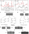
Mechanism of self-healing in A6ACA hydrogels. Raman (A) and FTIR–ATR (B) spectroscopy of healed (low pH) and unhealed (high pH) hydrogels demonstrating the presence of multiple types of hydrogen-bonded carboxyl groups. (C) Deduced molecular structures of pendant side chains in the face-on and interleaved hydrogen-bonding configurations responsible for the healing at low pH. (D) Structure of the pendant side chains in the unhealed hydrogels at high pH. At high pH, the carboxyl groups become deprotonated, leading to strong electrostatic repulsion between the apposing side chains, thus preventing healing. (E) Schematic explanation for why the healed hydrogels exhibit a mechanically stronger weld line compared to the bulk after healing for small timescales, and vice versa at very long times. Darker gray represents the toughened regions of the hydrogels due to protonation. The lighter gray represents the deprotonated (softer) regions of the hydrogels, which protonates and toughens with increasing exposure to low-pH solution.
Fig. 3.
Fig. 3.
Characterization of healing and healed hydrogels. (A) Effect of healing time on fracture stress. (B) Stress–strain curve, comparing tensile properties of 24-h healed gels with a single, unhealed hydrogel at identical conditions. The solid and dashed lines represent data from healed and unhealed hydrogels, respectively. (C) Fracture stress as a function of the extent of cross-linking for hydrogels containing 0.1%, 0.2%, and 0.5% of cross-linker (N, N′-methylenebisacrylamide) content. Error bars in A and C represent the standard deviation (n = 3).
Fig. 4.
Fig. 4.
Effect of side-chain length on the accessibility of functional groups. (A) Solubility of carboxylic acids of varying hydrocarbon chain lengths in water (black circles). Dashed red line indicates the density of carboxyl groups present in the hydrogels. (B) Molecular dynamics simulations setup for A6ACA network. A nine-arm motif of the network (Left) is used to create the 3D network structure (Right) via periodic boundary conditions. (C) Computed accessibilities of the amide and carboxyl groups in the A6ACA, A8ACA, and A11AUA hydrogels. (D) Representative configuration of the A6ACA and A11AUA network obtained from molecular dynamics simulations, shown in terms of solvent excluded surface, illustrating the higher accessibility of the amide groups in the former network. Blue, red, light gray, and white colors correspond to the surfaces of nitrogen, oxygen, carbon, and hydrogen, respectively. Chain length n in A and C represent number of CH2 groups in the carboxylic acids and side chains, respectively.
Fig. 5.
Fig. 5.
Applications of self-healing A6ACA hydrogels. The rupture site within the A6ACA coating on a polystyrene surface (A) before and (B) after healing. The coating was colored using a dye for easy visualization and the observed color change after healing is caused by its exposure to low-pH buffer. (Scale bars: 500 μm.) (C) Adhesion of A6ACA hydrogels to a poly(propylene) surface. (D) Polypropylene container holding acid solution after sealing the hole with A6ACA hydrogel. The arrow indicates the sealed site. (E) Adhesion of A6ACA hydrogels to rabbit gastric mucosa. (F) Cumulative tetracycline release from A6ACA hydrogels plotted as a function of time. Error bars represent standard deviation (n = 4).

References

    1. Varghese S, Lele AK, Mashelkar RA. Designing new thermoreversible gels by molecular tailoring of hydrophilic-hydrophobic interactions. J Chem Phys. 2000;112:3063–3070.
    1. Varghese S, Lele AK, Srinivas D, Sastry M, Mashelkar RA. Novel macroscopic self-organization in polymer gels. Adv Mater. 2001;13:1544–1548.
    1. Varghese S, Lele AK, Mashelkar R. Metal-ion-medaited healing of gels. J Polym Sci A1. 2006;44:666–670.
    1. Wojtecki RJ, Meador MA, Rowan SJ. Using the dynamic bond to access macroscopically responsive structurally dynamic polymers. Nat Mater. 2011;10:14–27. - PubMed
    1. Hager MD, Greil P, Leyens C, van der Zwaag S, Schubert US. Self-healing materials. Adv Mater. 2010;22:5424–5430. - PubMed

Publication types

LinkOut - more resources