. 2012 Mar 20;109(12):4592-7.
doi: 10.1073/pnas.1113748109. Epub 2012 Mar 5.

Transferred melanoma-specific CD8+ T cells persist, mediate tumor regression, and acquire central memory phenotype

Affiliations

Transferred melanoma-specific CD8+ T cells persist, mediate tumor regression, and acquire central memory phenotype

Aude G Chapuis et al. Proc Natl Acad Sci U S A. .

Abstract

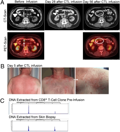
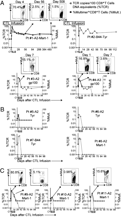
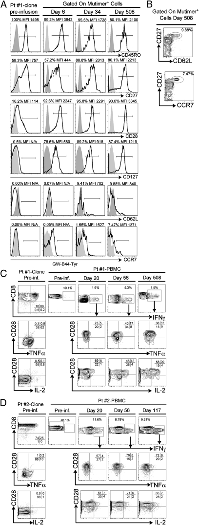
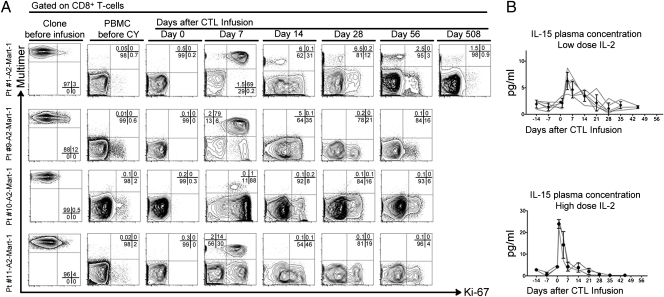
Adoptively transferred tumor-specific T cells offer the potential for non-cross-resistant therapy and long-term immunoprotection. Strategies to enhance in vivo persistence of transferred T cells can lead to improved antitumor efficacy. However, the extrinsic (patient conditioning) and intrinsic (effector cell) factors contributing to long-term in vivo persistence are not well-defined. As a means to enhance persistence of infused T cells in vivo and limit toxicity, 11 patients with refractory, progressive metastatic melanoma received cyclophosphamide alone as conditioning before the infusion of peripheral blood mononuclear cell-derived, antigen-specific, CD8(+) cytotoxic T-lymphocyte (CTL) clones followed by low-dose or high-dose IL-2. No life-threatening toxicities occurred with low-dose IL-2. Five of 10 evaluable patients had stable disease at 8 wk, and 1 of 11 had a complete remission that continued for longer than 3 y. On-target autoimmune events with the early appearance of skin rashes were observed in patients with stable disease or complete remission at 4 wk or longer. In vivo tracking revealed that the conditioning regimen provided a favorable milieu that enabled CTL proliferation early after transfer and localization to nonvascular compartments, such as skin and lymph nodes. CTL clones, on infusion, were characterized by an effector memory phenotype, and CTL that persisted long term acquired phenotypic and/or functional qualities of central memory type CTLs in vivo. The use of a T-cell product composed of a clonal population of antigen-specific CTLs afforded the opportunity to demonstrate phenotypic and/or functional conversion to a central memory type with the potential for sustained clinical benefit.


Conflict of interest statement

The authors declare no conflict of interest.

Figures

Fig. 1.
Fig. 1.
Complete response associated with a skin rash infiltrated with the adoptively transferred CTL clone. (A) Abdominal computed tomography (CT) (Upper) and PET scan combined with low-resolution noncontrast CT (Lower) before infusion (Left) and 28 d (Center) and 56 d after infusion (Right). Arrows indicate radiological image and active fluorodeoxyglucose (FDG) uptake before treatment, and circled areas show absence of detectable image and FDG uptake. (B) Back (Left), front chest (Center), and close-up of the front chest lesion (Right) of patient 1's rash 5 d after CTL infusion. (C) Superposition of TCR-γ length analysis of DNA extracted from the CTL clone preinfusion (Upper) and paraffin-embedded skin biopsy (Lower).
Fig. 2.
Fig. 2.
In vivo persistence of melanoma-specific CTL clones. (A) TCR copies per 100 CD8+ T-cell DNA equivalents (Left, x axis) and percentage of multimer-positive (%Mult.) CD8+ T cells (Right, x axis) in PBMCs collected 7 d (±2 d) before and after infusions for four of eight patients in cohort 1 whose clones showed persistence (≥48 d). Inset dot plots above the graph for patient 1 show percentages of multimer-positive CD8+ T cells at days 4, 56, and 508. (B) Same analysis performed for four of eight patients in cohort 1 whose clones demonstrated persistence for <7 d. (C) Plots for three patients in cohort 2. Inset dot plots show the percentages of multimer-positive CD8+ T cells in PBMCs at the time point peak frequencies observed. Pt, patient.
Fig. 3.
Fig. 3.
Adoptively transferred melanoma-specific CTL clones show sustained functional capacity in peripheral blood and tumor-infiltrated lymph nodes. (A) IFN-γ spot-forming cells per 105 PBMCs for patient (Pt) 1 (Left) and patient 2 (Right) after stimulation with unpulsed (empty circles) or HLA-A*0201-Mart1- or HLA-B*4402-tyrosinase-pulsed monocytes (filled circles), respectively, over time (y axis). (B) Stain of a cervical lymph node from patient 2 51 d after infusion stained with H&E (Left) and tyrosinase (Right). White arrows indicate lymphocyte infiltrates, and black arrows indicate invasive tyrosinase-positive melanoma. Images were collected with an objective with a magnification of 10×. (C) IFN-γ spot-forming cells per 105 cells (y axis). (Left to Right) Cells isolated from patient 2's infiltrated lymph node (LN) with unpulsed autologous monocytes (M); unpulsed M and infused clone cells; HLA-B*4402-tyrosinase-pulsed M only (negative controls); HLA-B*4402-tyrosinase-pulsed M and infused clone (positive control); and HLA-B*4402-tyrosinase-pulsed M and LN.
Fig. 4.
Fig. 4.
Adoptively transferred melanoma-specific CTLs show phenotypic and functional characteristics shared with CD8+ Tcm in vivo. (A) Patient (Pt) 1: Expression of CD45RO, CD27, CD28, CD127 CD62L, and CCR7 (bold lines) on the HLA*0201-Mart1-specific CTL clone preinfusion (Pre-inf.; Left), and on days 6, 34, and 508 after transfer (Right). MFIs are shown for surface markers with >1% positivity compared with isotype controls (gray areas). (B) Expression of CD27 (y axis), CD62L (x axis) (Upper), and CCR7 (Lower) gated on multimer-positive CTLs 508 d after transfer. Expression of CD28; secretion of TNF-α and IL-2 within IFN-γ+ cells on infused clones; and PBMCs preinfusion and on days 20, 56, and 508 (C; patient 1) and days 20, 56, and 117 (D; patient 2).
Fig. 5.
Fig. 5.
CY conditioning followed by exogenous IL-2 fosters a favorable environment for replication of infused CTLs. (A) Intranuclear Ki-67 expression on clones (Left) and CD8+ multimer-positive or -negative cells within PBMCs of patients 1, 9, 10, and 11 (rows) before CY and on days 0, 7, 14, 28, 56, and 508 (patient 1 only) after T-cell infusion (columns). Multimer (y axis) and Ki-67 (x axis). (B) IL-15 plasma levels (pg/mL) (y axis) plotted over time (x axis). Gray lines represent individual patients, and filled circles show mean and SD. Patients in cohort 1 (Upper) and cohort 2 (Lower).

References

    1. Hunder NN, et al. Treatment of metastatic melanoma with autologous CD4+ T cells against NY-ESO-1. N Engl J Med. 2008;358:2698–2703. - PMC - PubMed
    1. Rosenberg SA, Dudley ME. Adoptive cell therapy for the treatment of patients with metastatic melanoma. Curr Opin Immunol. 2009;21:233–240. - PMC - PubMed
    1. Yee C, et al. Adoptive T cell therapy using antigen-specific CD8+ T cell clones for the treatment of patients with metastatic melanoma: In vivo persistence, migration, and antitumor effect of transferred T cells. Proc Natl Acad Sci USA. 2002;99:16168–16173. - PMC - PubMed
    1. Butler MO, et al. Establishment of antitumor memory in humans using in vitro-educated CD8+ T cells. Sci Transl Med. 2011 3:80ra24. - PMC - PubMed
    1. Riddell SR, et al. Restoration of viral immunity in immunodeficient humans by the adoptive transfer of T cell clones. Science. 1992;257:238–241. - PubMed

Publication types

MeSH terms