. 2012 Mar 20;109(12):4690-5.
doi: 10.1073/pnas.1113455109. Epub 2012 Mar 5.

Salience network integrity predicts default mode network function after traumatic brain injury

Affiliations

Salience network integrity predicts default mode network function after traumatic brain injury

Valerie Bonnelle et al. Proc Natl Acad Sci U S A. .

Abstract

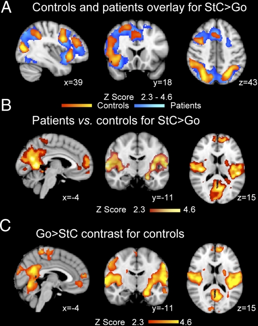
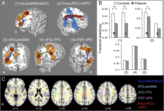
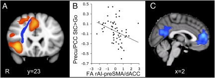
Efficient behavior involves the coordinated activity of large-scale brain networks, but the way in which these networks interact is uncertain. One theory is that the salience network (SN)--which includes the anterior cingulate cortex, presupplementary motor area, and anterior insulae--regulates dynamic changes in other networks. If this is the case, then damage to the structural connectivity of the SN should disrupt the regulation of associated networks. To investigate this hypothesis, we studied a group of 57 patients with cognitive impairments following traumatic brain injury (TBI) and 25 control subjects using the stop-signal task. The pattern of brain activity associated with stop-signal task performance was studied by using functional MRI, and the structural integrity of network connections was quantified by using diffusion tensor imaging. Efficient inhibitory control was associated with rapid deactivation within parts of the default mode network (DMN), including the precuneus and posterior cingulate cortex. TBI patients showed a failure of DMN deactivation, which was associated with an impairment of inhibitory control. TBI frequently results in traumatic axonal injury, which can disconnect brain networks by damaging white matter tracts. The abnormality of DMN function was specifically predicted by the amount of white matter damage in the SN tract connecting the right anterior insulae to the presupplementary motor area and dorsal anterior cingulate cortex. The results provide evidence that structural integrity of the SN is necessary for the efficient regulation of activity in the DMN, and that a failure of this regulation leads to inefficient cognitive control.


Conflict of interest statement

The authors declare no conflict of interest.

Figures

Fig. 1.
Fig. 1.
Abnormal DMN deactivation after TBI. (A) Overlay of brain activation associated with correct stop (StC) vs. go trials for patients (blue) and controls (yellow-red). (B) Brain regions where patients show greater brain activation for StC > Go than controls. (C) Brain regions showing greater activation for go than stop trials in the control group. Results are superimposed on the MNI 152 T1 1-mm brain template. Cluster corrected Z = 2.3, P < 0.05.
Fig. 2.
Fig. 2.
Comparison of white matter structure between patients and controls. (A and C) White matter tracts of interest are represented in 3D on the MNI-152 T1 1-mm brain 3D template (A) and in 2D on axial MNI-152 T1 1-mm brain views, overlaid on tract-based spatial statistics (TBSS) white matter skeleton (yellow) (C). (A) The resulting probabilistic tractography tracts are shown for the connections between the rAI and the preSMA/dACC (dark blue) (1), the precu/PCC and vmPFC bilaterally (red) (2), the rIFG and the preSMA (light blue) (3), the rIFG and the right TPJ (rTPJ) (green; 4) and the rFEF and rIPS (lilac/pink; 5). Orange and blue areas represent the mean BOLD signal change in patients and controls for the contrasts StC > Go and Go > StC, respectively. (B) The bar charts show FA ± SEM within each tract compared between TBI patients (gray) and 30 age-matched controls (white). R, right; L, left. *P < 0.05; **P < 0.005.
Fig. 3.
Fig. 3.
The integrity of the rAI–preSMA/dACC white matter tract predicts DMN deactivation. (A) Coronal view of the white matter connection between rAI and preSMA/dACC (blue) overlaid on the activation map for the contrast StC > Go in patients (orange), superimposed on the MNI 152 T1 1-mm brain template. (B) FA of the rAI–preSMA/dACC tracts in patients plotted against the percent signal change within the precu/PCC ROI on correct stop trials relative to go trials. FA measures are corrected for age and whole-brain mean FA and normalized. (C) Sagittal view of the brain regions showing negatively correlated activation with FA measures within the rAI–preSMA/dACC tract for StC relative to go trials, superimposed on the MNI 152 T1 2-mm brain template. Cluster corrected Z = 2.3, P < 0.05.

References

    1. Bressler SL, Menon V. Large-scale brain networks in cognition: Emerging methods and principles. Trends Cogn Sci. 2010;14:277–290. - PubMed
    1. Mesulam M. Defining neurocognitive networks in the BOLD new world of computed connectivity. Neuron. 2009;62:1–3. - PubMed
    1. Damoiseaux JS, et al. Consistent resting-state networks across healthy subjects. Proc Natl Acad Sci USA. 2006;103:13848–13853. - PMC - PubMed
    1. Seeley WW, et al. Dissociable intrinsic connectivity networks for salience processing and executive control. J Neurosci. 2007;27:2349–2356. - PMC - PubMed
    1. Beckmann CF, DeLuca M, Devlin JT, Smith SM. Investigations into resting-state connectivity using independent component analysis. Philos Trans R Soc Lond B Biol Sci. 2005;360:1001–1013. - PMC - PubMed

Publication types

LinkOut - more resources