. 2012 Mar 12;209(3):471-8.
doi: 10.1084/jem.20112401. Epub 2012 Mar 5.

Primary Epstein-Barr virus infection does not erode preexisting CD8⁺ T cell memory in humans

Affiliations

Primary Epstein-Barr virus infection does not erode preexisting CD8⁺ T cell memory in humans

Oludare A Odumade et al. J Exp Med. .

Abstract

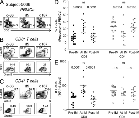
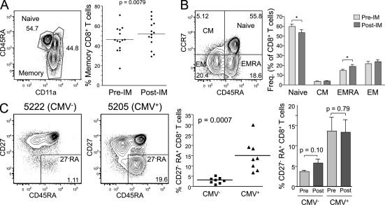
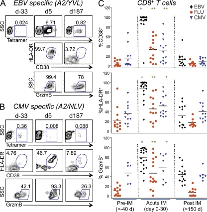
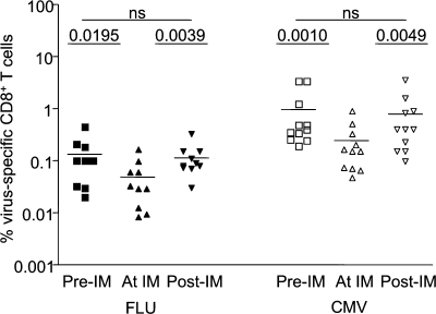
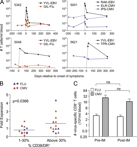
Acute Epstein-Barr virus (EBV) infection results in an unusually robust CD8(+) T cell response in young adults. Based on mouse studies, such a response would be predicted to result in attrition of preexisting memory to heterologous infections like influenza A (Flu) and cytomegalovirus (CMV). Furthermore, many studies have attempted to define the lymphocytosis that occurs during acute EBV infection in humans, but it is unclear whether bystander T cells contribute to it. To address these issues, we performed a longitudinal prospective study of primary EBV infection in humans. During acute EBV infection, both preexisting CMV- and Flu-specific memory CD8(+) T cells showed signs of bystander activation, including up-regulation of granzyme B. However, they generally did not expand, suggesting that the profound CD8(+) lymphocytosis associated with acute EBV infection is composed largely of EBV-specific T cells. Importantly, the numbers of CMV- and Flu-specific T cells were comparable before and after acute EBV infection. The data support the concept that, in humans, a robust CD8(+) T cell response creates a new memory CD8(+) T cell niche without substantially depleting preexisting memory for heterologous infections.


Figures

Figure 1.
Figure 1.
Extensive CD8+ T cell activation and expansion occur during primary EBV infection. PBMCs from each subject were stained with antibodies against CD3, CD4, CD8, CD38, CD45RA, DR, and GrzmB. Panels A–C show sample flow cytometric analysis from subject 5036 before, during, and after acute infection with EBV. d, day relative to symptom onset. (A) CD4 and CD8 expression on CD3+ T cells over time. (B and C) Dot plots showing activation markers CD38, HLA-DR (DR), or GrzmB expression on total CD3+CD8+ T cells (B) or total CD3+CD4+ T cells (C). (D and E) The frequency (D) or number (E) of CD8+ or CD4+ T cells before (Pre-IM), during (At IM), and after (Post-IM) acute primary EBV infection (infectious mononucleosis) is shown. Wilcoxon signed rank test, P < 0.05 considered statistically significant (n = 14). Horizontal bars indicate the mean.
Figure 2.
Figure 2.
Primary EBV infection results in a slight increase in effector memory RA+, but not CD27RA+, CD8+ T cells. PBMCs from each subject were stained with antibodies against CD3, CD4, CD8, CD11a, CD45RA, CCR7, CD62L, and CD27. All panels show analysis of CD3+CD8+ T cells. (A) Memory phenotype CD8+ T cells were defined as CD11ahi. The percentage of cells in this gate was analyzed in 14 subjects before (Pre-IM) or at least 350 d after primary EBV infection (Post-IM). The mean day after infection was 750 (the p-value was determined using a paired Student’s t test). (B) CCR7 and CD45RA expression was examined on CD8+ T cells from subjects before or at least 350 d after primary EBV infection. Central memory (CM), effector memory (EM), and effector memory RA (EMRA) subsets were defined as shown. Statistically significant changes are indicated with an asterisk (paired Student’s t test: *, P < 0.05). Error bars indicate standard deviation. (C) CD27 and CD45RA expression was examined on CD8+ T cells in 16 subjects. Eight of these were CMV seropositive at enrollment, and the other eight were CMV seronegative and remained negative during the duration of the study. Scatter plot compares the percentage of CD27 negative, CD45RA+ (CD27RA) CD8+ T cells in CMV seropositive or seronegative individuals (n = 8/group; unpaired Student’s t test, significant). The bar graph shows the mean and standard deviation of CD27RA CD8+ T cells before versus after primary EBV infection (n = 6/group; paired Student’s t test, not significant for either). (A and C) Horizontal bars indicate the mean.
Figure 3.
Figure 3.
Both EBV-specific and bystander (CMV and Flu specific) CD8+ T cells were activated during acute EBV infection. PBMCs from each subject were stained with antibodies against CD3, CD4, CD8, CD38, CD45RA, DR, and GrzmB, as well as pMHCI tetramers. Panels A and B show sample flow cytometric analysis of PBMCs from subject 5036 before, during, and after acute infection with EBV. (A) HLA-A2/YVL (EBV) tetramer staining on CD8+ T cells. (B) HLA-A2/NLV (CMV) tetramer staining on CD8+ T cells. (A and B) Lower dot plots show CD38, HLA-DR (DR), and GrzmB staining of tetramer+ cells. (C) Percentage of tetramer+CD8+ T cells that expressed CD38 (top), DR (middle), or GrzmB (bottom) before (>40 d prior), during (0–30 d), and after (>150 d) acute primary EBV infection. EBV-specific CD8+ T cells were identified with HLA-A2/YVL, A3/RVR, or B8/RAK tetramers; Flu-specific CD8+ T cells were identified with HLA-A2/GIL tetramers; and CMV-specific CD8+ T cells were identified with HLA-A2/NLV, B7/TPR, B8/ELR, or B35/IPS tetramers (n = 6–19 per virus-specific T cell; ANOVA: *, P < 0.05; **, P < 0.001). Horizontal bars indicate the mean.
Figure 4.
Figure 4.
There is a transient reduction in the frequency of Flu- and CMV-specific T cells during primary EBV infection. PBMCs were stained with antibodies against CD3, CD4, CD8, and pMHCI tetramers. The graph represents the frequency of Flu (left) and CMV (right) tetramer+CD3+CD8+ T cells before (squares), during (triangles), and after (inverted triangles) acute primary EBV infection (n = 10 for Flu and n = 11 for CMV; paired Student’s t test, P > 0.05 was considered not significant). Horizontal bars indicate the mean.
Figure 5.
Figure 5.
There is no attrition of preexisting memory CD8+ T cells after primary EBV infection. (A) Panels show data from four individual subjects depicting the total number of CD8+ T cells per milliliter of blood that stained with the indicated tetramers for EBV (black diamond), Flu (red circle), or CMV (blue inverted triangle) over time. Dotted lines indicate that EBV-specific T cells were not detectable before primary infection. (B) The fold expansion of CMV- and Flu-specific CD8+ T cells in subjects that displayed a more activated phenotype (based on >30% expression of CD38 and DR) or less activated phenotype (1–30% expression of CD38 and DR). Student’s t test, P < 0.05 (n = 26). Horizontal bars indicate the mean. (C) Flu- and CMV-specific CD8+ T cell numbers before and >150 d after primary EBV infection. Paired Student’s t test, P > 0.05 (n = 10 for Flu and n = 11 for CMV). Error bars indicate standard deviation.

References

    1. Bahl K., Kim S.K., Calcagno C., Ghersi D., Puzone R., Celada F., Selin L.K., Welsh R.M. 2006. IFN-induced attrition of CD8 T cells in the presence or absence of cognate antigen during the early stages of viral infections. J. Immunol. 176:4284–4295 - PubMed
    1. Balfour H.H., Jr, Holman C.J., Hokanson K.M., Lelonek M.M., Giesbrecht J.E., White D.R., Schmeling D.O., Webb C.H., Cavert W., Wang D.H., Brundage R.C. 2005. A prospective clinical study of Epstein-Barr virus and host interactions during acute infectious mononucleosis. J. Infect. Dis. 192:1505–1512 10.1086/491740 - DOI - PubMed
    1. Biglino A., Sinicco A., Forno B., Pollono A.M., Sciandra M., Martini C., Pich P., Gioannini P. 1996. Serum cytokine profiles in acute primary HIV-1 infection and in infectious mononucleosis. Clin. Immunol. Immunopathol. 78:61–69 10.1006/clin.1996.0009 - DOI - PubMed
    1. Callan M.F., Tan L., Annels N., Ogg G.S., Wilson J.D., O’Callaghan C.A., Steven N., McMichael A.J., Rickinson A.B. 1998. Direct visualization of antigen-specific CD8+ T cells during the primary immune response to Epstein-Barr virus in vivo. J. Exp. Med. 187:1395–1402 10.1084/jem.187.9.1395 - DOI - PMC - PubMed
    1. Clute S.C., Watkin L.B., Cornberg M., Naumov Y.N., Sullivan J.L., Luzuriaga K., Welsh R.M., Selin L.K. 2005. Cross-reactive influenza virus-specific CD8+ T cells contribute to lymphoproliferation in Epstein-Barr virus-associated infectious mononucleosis. J. Clin. Invest. 115:3602–3612 10.1172/JCI25078 - DOI - PMC - PubMed

Publication types

MeSH terms