. 2012 Jun 1;125(Pt 11):2732-9.
doi: 10.1242/jcs.100909. Epub 2012 Mar 5.

A RUNX2-HDAC1 co-repressor complex regulates rRNA gene expression by modulating UBF acetylation

Affiliations

A RUNX2-HDAC1 co-repressor complex regulates rRNA gene expression by modulating UBF acetylation

Syed A Ali et al. J Cell Sci. .

Abstract

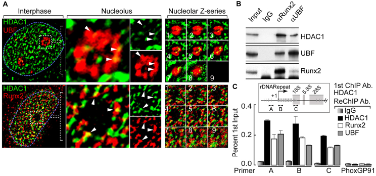
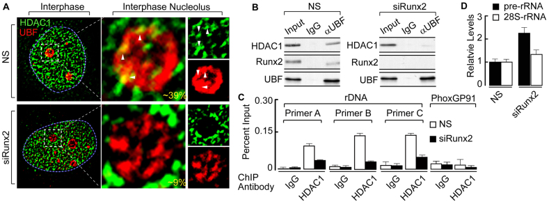
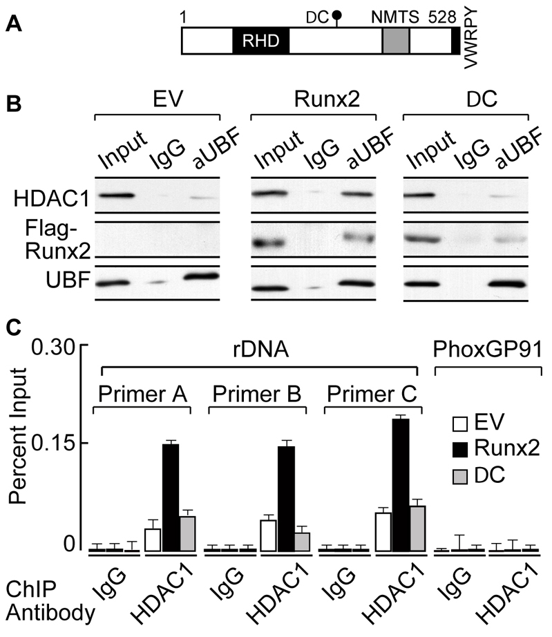
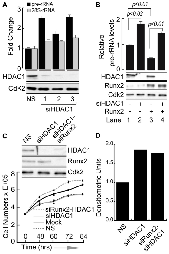
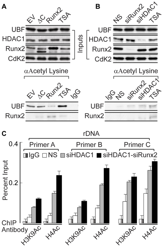
The osteogenic and oncogenic transcription factor RUNX2 downregulates the RNA polymerase I (RNA Pol I)-mediated transcription of rRNAs and changes histone modifications associated with the rDNA repeat. However, the mechanisms by which RUNX2 suppresses rRNA transcription are not well understood. RUNX2 cofactors such as histone deacetylases (HDACs) play a key role in chromatin remodeling and regulation of gene transcription. Here, we show that RUNX2 recruits HDAC1 to the rDNA repeats in osseous cells. This recruitment alters the histone modifications associated with active rRNA-encoding genes and causes deacetylation of the protein upstream binding factor (UBF, also known as UBTF). Downregulation of RUNX2 expression reduces the localization of HDAC1 to the nucleolar periphery and also decreases the association between HDAC1 and UBF. Functionally, depletion of HDAC1 relieves the RUNX2-mediated repression of rRNA-encoding genes and concomitantly increases cell proliferation and global protein synthesis in osseous cells. Our findings collectively identify a RUNX2-HDAC1-dependent mechanism for the regulation of rRNA-encoding genes and suggest that there is plasticity to RUNX2-mediated epigenetic control, which is mediated through selective mitotic exclusion of co-regulatory factors.


Figures

Fig. 1.
Fig. 1.
HDAC1 colocalizes with RUNX2 and UBF in the interphase nucleolus. (A) Immunofluorescence microscopy in SaOS-2 cells demonstrates HDAC1 colocalization with UBF (upper panels) and RUNX2 (lower panels) at the nucleolar periphery. The blue dotted line represents the nucleus and the white dotted box marks a nucleolus. (B) Co-immunoprecipitation assays show that HDAC1 associates with UBF and RUNX2 in proliferating osteosarcoma cells. (C) ChIP-ReChIP experiments show the binding of HDAC1 together with RUNX2 and UBF at rDNA repeats (primer A, B and C). Primary (1st) ChIP was performed using anti-HDAC1 antibody (Ab) followed by ReChIP (2nd) using anti-HDAC1, anti-RUNX2 and anti-UBF antibodies. A Phox-GP91 gene primer was used as a negative control to demonstrate the specificity of the ChIP-ReChIP assays. In the inset, a single rDNA repeat is shown with the location of the primer sets used in ChIP assays (A, B and C) at different regions of the rDNA. Vertical lines indicate the RUNX2-binding sites at the rDNA repeat. White arrows show the location of primers.
Fig. 2.
Fig. 2.
Decrease in HDAC1 nucleolar recruitment positively regulates rRNA gene expression. (A) Knockdown of RUNX2 by using siRNA oligonucleotides decreases HDAC1 recruitment at the periphery of interphase nucleolus (~9%), when compared with non-specific (NS) control (~39%) in SaOS-2 cells as analyzed by immunofluorescence microscopy. The blue dotted line represents the nucleus and the white dotted box marks an interphase nucleolus. The percentage reflects the proportion of the total nucleoli (randomly selected for analysis) that show colocalization between HDAC1 and UBF at the nucleolus. (B) Immunoprecipitation experiments showing that reduction in RUNX2 levels decreases UBF association with HDAC1 as compared with non-specific control sample. (C) ChIP assays demonstrating that knocking down RUNX2 decreases HDAC1 rDNA binding, as shown by rDNA primer sets A, B and C. HDAC1 shows no binding to the unrelated negative control gene Phox-GP91. (D) RUNX2 knockdown, concomitant with decreased UBF association with HDAC1 and reduced HDAC1 occupancy of rDNA, also decreases the pre-rRNA levels as determined by RT-qPCR. To normalize the values β-actin-encoding mRNA was used as an internal control.
Fig. 3.
Fig. 3.
The RUNX2 C-terminus mediates HDAC1 association with UBF and rDNA. (A) A schematic representation of full-length RUNX2 protein indicating the Runt homology domain (RHD) for DNA binding, the nuclear-matrix-targeting signal (NMTS) and the C-terminus, showing the mark for ΔC truncation protein (DC). (B) Immunoprecipitation analysis demonstrates that overexpression of WT-RUNX2, increases UBF association with HDAC1 but not the ΔC mutant (DC), when compared with experiments using the empty vector (EV) control in osteosarcoma cells. (C) To analyze RUNX2-mediated rDNA occupancy of HDAC1, either full-length RUNX2 or the ΔC mutant was expressed in SaOS-2 cells. A ChIP assay shows that overexpression of WT-RUNX2, but not the ΔC mutant, increases HDAC1 occupancy at rDNA, as determined by primer set A, B and C. RUNX2 (WT and ΔC) does not bind to the Phox-GP91 gene, demonstrating the specificity of ChIP experiments.
Fig. 4.
Fig. 4.
Combined knockdown of HDAC1 and RUNX2 increases cell proliferation and the overall protein synthesis rate. (A) Western blot showing the knockdown of HDAC1 protein by three independent siRNA oligonucleotides after 48 hours. The bar graph demonstrates the increased expression of rRNA genes in SaOS-2 cells as assessed by RT-qPCR. (B) Western blot showing siHDAC1 (lane 2), WT-RUNX2 overexpression (lane 3) and siHDAC1 with RUNX2 overexpression (lane 4) as compared with control (lane 1). In parallel samples, the effect on pre-rRNA expression was determined by RT-qPCR, using β-actin-encoding mRNA as an internal control (bar graphs). (C) Western blot showing the effect of siHDAC1 alone or siHDAC1 in combination with siRUNX2 after 48 hours of treatment with specific oligonucleotides. The line graph represents the increased cell count when either HDAC1 alone or together with RUNX2 was downregulated in SaOS-2 cells. (D) The bar graph shows combined densitometric quantification (Image J) of radioactive proteins from cells metabolically labeled with [35S]methionine. Each bar represents results from two independent experiments in which either HDAC1 alone was depleted or both HDAC1 and RUNX2 were targeted with siRNAs. NS, non-silencing siRNA.
Fig. 5.
Fig. 5.
Deacetylation of UBF requires RUNX2-mediated HDAC1 recruitment. (A) Western blots showing the effect of expression of wild-type RUNX2 and the ΔC mutant after 48 hours of lentiviral infection in SaOS-2 cells. Samples were also treated with the histone deacetylase inhibitor TSA as control (upper panel). An immunoprecipitation assay was performed using an antibody against acetylated lysine, demonstrating decreased UBF acetylation in the presence of wild-type RUNX2 but not the ΔC mutant when compared with empty vector (EV) controls (lower panel). (B) Western blots demonstrating the depletion of RUNX2 and HDAC1 by siRNA oligonucleotides and treatment with TSA (upper panel). An antibody against acetylated lysine was used for immunoprecipitation. There is an increase in UBF acetylation in siRUNX2, siHDAC1 and TSA-treated samples, when compared with the non-silencing (NS) control (lower panel). (C) ChIP analysis showing that there is an increase in active histone marks (H3K9Ac and H4Ac) associated with rDNA, in the absence of HDAC1 alone or upon combined depletion of HDAC1 and RUNX2 after 48 hours of siRNA oligonucleotide treatment, as assessed by use of rDNA primer A, B and C.

References

    1. Ali S. A., Zaidi S. K., Dacwag C. S., Salma N., Young D. W., Shakoori A. R., Montecino M. A., Lian J. B., van Wijnen A. J., Imbalzano A. N., et al. (2008). Phenotypic transcription factors epigenetically mediate cell growth control. Proc. Natl. Acad. Sci. USA 105, 6632-6637 - PMC - PubMed
    1. Ali S. A., Zaidi S. K., Dobson J. R., Shakoori A. R., Lian J. B., Stein J. L., van Wijnen A. J., Stein G. S. (2010). Transcriptional corepressor TLE1 functions with Runx2 in epigenetic repression of ribosomal RNA genes. Proc. Natl. Acad. Sci. USA 107, 4165-4169 - PMC - PubMed
    1. Arabi A., Wu S., Ridderstråle K., Bierhoff H., Shiue C., Fatyol K., Fahlén S., Hydbring P., Söderberg O., Grummt I., et al. (2005). c-Myc associates with ribosomal DNA and activates RNA polymerase I transcription. Nat. Cell Biol. 7, 303-310 - PubMed
    1. Bell S. P., Learned R. M., Jantzen H. M., Tjian R. (1988). Functional cooperativity between transcription factors UBF1 and SL1 mediates human ribosomal RNA synthesis. Science 241, 1192-1197 - PubMed
    1. Budde A., Grummt I. (1999). p53 represses ribosomal gene transcription. Oncogene 18, 1119-1124 - PubMed

Publication types

MeSH terms