. 2012;7(2):e32157.
doi: 10.1371/journal.pone.0032157. Epub 2012 Feb 29.

Lethal Nipah virus infection induces rapid overexpression of CXCL10

Affiliations

Lethal Nipah virus infection induces rapid overexpression of CXCL10

Cyrille Mathieu et al. PLoS One. 2012.

Abstract

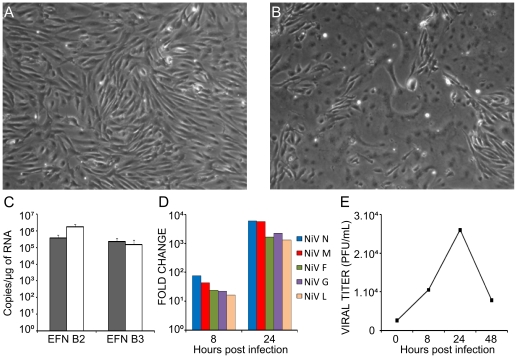
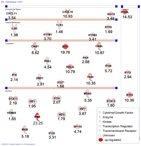
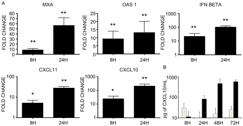
Nipah virus (NiV) is a recently emerged zoonotic Paramyxovirus that causes regular outbreaks in East Asia with mortality rate exceeding 75%. Major cellular targets of NiV infection are endothelial cells and neurons. To better understand virus-host interaction, we analyzed the transcriptome profile of NiV infection in primary human umbilical vein endothelial cells. We further assessed some of the obtained results by in vitro and in vivo methods in a hamster model and in brain samples from NiV-infected patients. We found that NiV infection strongly induces genes involved in interferon response in endothelial cells. Among the top ten upregulated genes, we identified the chemokine CXCL10 (interferon-induced protein 10, IP-10), an important chemoattractant involved in the generation of inflammatory immune response and neurotoxicity. In NiV-infected hamsters, which develop pathology similar to what is seen in humans, expression of CXCL10 mRNA was induced in different organs with kinetics that followed NiV replication. Finally, we showed intense staining for CXCL10 in the brain of patients who succumbed to lethal NiV infection during the outbreak in Malaysia, confirming induction of this chemokine in fatal human infections. This study sheds new light on NiV pathogenesis, indicating the role of CXCL10 during the course of infection and suggests that this chemokine may serve as a potential new marker for lethal NiV encephalitis.


Conflict of interest statement

Competing Interests: The authors have declared that no competing interests exist.

Figures

Figure 1
Figure 1. Primary human endothelial cells are highly permissive to NiV-infection.
A, Mock-infected and B, NiV-infected HUVECs (MOI = 1) observed 24 h after infection show extensively developed syncytium formation. C, RT-qPCR analysis of the expression of NiV receptors ephrinB2 (EFNB2) and ephrinB3 (EFNB3) in HUVECs (black bars) and U373 astroglioma cells (white bars). D, Production of RNA specific for NiV genes: nucleoprotein (N), matrix (M), fusion protein (F), glycoprotein (G) and polymerase (L) in HUVECs during infection, determined by RT-qPCR. Fold change is relative to the number of copies of viral mRNAs 8 h or 24 h post infection compared to the number of copies obtained after 1 h of contact with the virus. E, Production of infectious NiV particles during the course of infection; supernatants taken at different time points were titrated on a Vero cell monolayer.
Figure 2
Figure 2. Impact of NiV infection on interferon pathway in HUVEC cells, determined using Ingenuity Pathway Analysis.
NiV-induced upregulated genes are presented grouped, depending the cell compartment in which the corresponding proteins a localized (nucleus, cytoplasm, plasma membrane and extracellular space, for secreted proteins). Level of FC increase is indicated bellow each gene and intensity of red color corresponds to the FC.
Figure 3
Figure 3. Analysis of expression of interferon-related genes in NiV-infected HUVECs.
A, RT-qPCR analysis of the expression of MXA, OAS1, IFN beta, CXCL11 and CXCL10. B, ELISA analysis of the production of CXCL10 protein in mock-infected (white bars) and NiV-infected (black bars) HUVC cultures. Results are expressed as the average of 2–3 individual experiments and bars represent standard deviation. *p<0.05, **p<0.01, Mann-Whitney U-test.
Figure 4
Figure 4. Expression of CXCL10 and NiV nucleoprotein (N) genes in organs obtained from NiV-infected hamsters during the course of infection.
Hamsters were sacrificed at different time points after infection and RNA was isolated from different organs and analyzed by RT-qPCR. Grey bars correspond to CXCL10 and black bars to NiV-N, calculated as described in Methods. Significant correlation was found between expression of CXCL10 and expression of NiV N (R2 = 0,989, p<0,001, Pearson test).
Figure 5
Figure 5. Immunohistochemical analysis of CXCL10 production in the brain durng NiV infection.
Non-infected patients (A, ×100) and patients autopsied after lethal acute NiV-infection (B–H) were analysed for the expression of CXCL10 in endothelial cells in cerebral cortex (B, D, ×1000, arrows) and cells with neuronal morphology (E, F). CXCL10 immunoreactivity in perivascular inflammatory cells (C, G, H, ×400).

References

    1. Eaton BT, Broder CC, Middleton D, Wang LF. Hendra and Nipah viruses: different and dangerous. Nat Rev Microbiol. 2006;4:23–35. - PMC - PubMed
    1. Chua KB, Bellini WJ, Rota PA, Harcourt BH, Tamin A, et al. Nipah virus: a recently emergent deadly paramyxovirus. Science. 2000;288:1432–1435. - PubMed
    1. Stone R. Epidemiology Breaking the Chain in Bangladesh. Science. 2011;331:1128–1131. - PubMed
    1. Luby PS, Hossain MJ, Gurley ES, Ahmed BN, Banu S, et al. Recurrent Zoonotic Transmission of Nipah Virus into Humans, Bangladesh, 2001–2007. Emerging Infectious Diseases. 2009;15:1229–1233. - PMC - PubMed
    1. Luby SP, Gurley ES, Hossain MJ. Transmission of human infection with Nipah virus. Clin Infect Dis. 2009;49:1743–1748. - PMC - PubMed

Publication types

MeSH terms