. 2012;7(2):e32524.
doi: 10.1371/journal.pone.0032524. Epub 2012 Feb 29.

Roles of the amino terminal region and repeat region of the Plasmodium berghei circumsporozoite protein in parasite infectivity

Affiliations

Roles of the amino terminal region and repeat region of the Plasmodium berghei circumsporozoite protein in parasite infectivity

Cassandra Aldrich et al. PLoS One. 2012.

Abstract

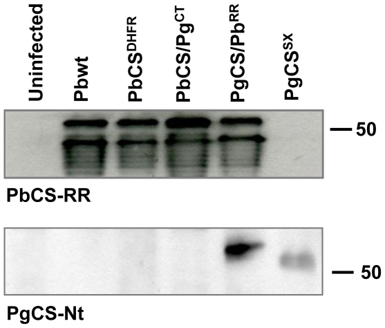
The circumsporozoite protein (CSP) plays a key role in malaria sporozoite infection of both mosquito salivary glands and the vertebrate host. The conserved Regions I and II have been well studied but little is known about the immunogenic central repeat region and the N-terminal region of the protein. Rodent malaria Plasmodium berghei parasites, in which the endogenous CS gene has been replaced with the avian Plasmodium gallinaceum CS (PgCS) sequence, develop normally in the A. stephensi mosquito midgut but the sporozoites are not infectious. We therefore generated P. berghei transgenic parasites carrying the PgCS gene, in which the repeat region was replaced with the homologous region of P. berghei CS (PbCS). A further line, in which both the N-terminal region and repeat region were replaced with the homologous regions of PbCS, was also generated. Introduction of the PbCS repeat region alone, into the PgCS gene, did not rescue sporozoite species-specific infectivity. However, the introduction of both the PbCS repeat region and the N-terminal region into the PgCS gene completely rescued infectivity, in both the mosquito vector and the mammalian host. Immunofluorescence experiments and western blot analysis revealed correct localization and proteolytic processing of CSP in the chimeric parasites. The results demonstrate, in vivo, that the repeat region of P. berghei CSP, alone, is unable to mediate sporozoite infectivity in either the mosquito or the mammalian host, but suggest an important role for the N-terminal region in sporozoite host cell invasion.


Conflict of interest statement

Competing Interests: The authors have declared that no competing interests exist.

Figures

Figure 1
Figure 1. Schematic representation and alignment of wild type and transgenic P.berghei circumsporozoite proteins.
(A) Schematic representation of the CS proteins present in each of the four transgenic P. berghei parasite lines generated. The corresponding names of the transgenic parasite lines are shown on the right. PbCSDHFR parasites carry the wildtype PbCS coding sequence (white boxes, RI and RII indicated with light and dark green respectively) and, similar to all transgenic lines, contain the T. gondii DHFR drug selectable marker inserted in the CS locus. PgCSSX parasites carry the full PgCS coding sequence (grey boxes, RI and RII indicated with light and dark blue respectively), but with the SpeI (S) and XhoI (X) restriction endonuclease sites inserted on either side of the repeat region. PgCS/PbRR parasites contain the PgCS N-terminal and C-terminal regions (grey boxes) and the PbCS repeat region (white box). PbCS/PgCT parasites carry the PbCS N-terminal and repeat regions (white boxes) and the PgCS C-terminal region (grey box). (B) Alignment of the wild type PbCS and transgenic PgCSSX and PgCS/PbRR amino acid sequences. Shaded boxes represent areas of amino acid identity. Regions I and II (black lines), the repeat region (blue line) and the SpeI and XhoI restriction sites (red lines) are labelled. The SpeI and XhoI sites were introduced into the PgCS sequence to mediate exchange of the PgCS repeat region with the PbCS repeat region.
Figure 2
Figure 2. Generation and southern blot analysis of transgenic PgCS/PbRR parasite lines.
(A) Schematic representation of (i) the PgCS/PbRR targeting construct, (ii) the wt PbCS locus and (iii) the targeted locus after recombination between the PbCS 5′UTR and 3′UTR sequences. The vertically dashed box indicates the 1.13 kb 5′UTR sequence used in the construct and the horizontally dashed boxes indicate the 0.3 kb and 0.85 kb 3′UTR sequences between which the TgDHFR-TS selectable marker cassette (light grey) was inserted in the construct. The PgCS/PbRR chimeric gene contains the PgCS N- and C-terminal regions (dark grey boxes) and the PbCS repeat region (white box) flanked by the SpeI (S) and XhoI (X) sites. In the wt PbCS locus the white box indicates the full PbCS gene. Thick black lines indicate the probes used in southern blots. E: EcoRV site. (B) Southern blot of EcoRV/SpeI (E/S) and EcoRV/XhoI (E/X) digested genomic DNA from Pbwt parasites and transgenic PgCS/PbRR parasites (clones 4 and 5), hybridised with the PbCS 3′UTR probe. Two bands of 4.0 and 1.5 kb were present in Pbwt DNA in both digestions. E/S digested DNA from the PgCS/PbRR clones revealed two bands of 1.1 and 2.0 kb, while E/X digested DNA revealed two bands of 0.6 and 2.0 kb, demonstrating the replacement of the endogenous PbCS gene with the targeted construct. (C) The same membrane was then hybridised with the PgCS N-terminal probe (PgCS-Nt), encompassing the entire N-terminal region of PgCS. No band was present in Pbwt DNA. However, a band of 3.1 kb or 3.6 kb was present in the E/S or E/X digested transgenic DNA, respectively.
Figure 3
Figure 3. Generation and southern blot analysis of transgenic PbCS/PgCT parasite lines.
(A) Schematic representation of (i) the PgCS/PbRR targeting construct, (ii) the wt PbCS locus and (iii) the targeted locus after recombination between the PbCS repeat region (white box) and the PbCS 3′UTR (horizontally dashed box). The PbCS/PgCT chimeric gene contains both the PbCS N- terminal and repeat regions (white boxes) and the PgCS C-terminal region (dark grey box). The light grey box indicates the TgDHFR-TS selectable marker cassette. Thick black lines indicate the probes used in southern blots. E = EcoRV site, S = SpeI site and X = XhoI site. (B) Southern blot of EcoRV/SpeI (E/S) and EcoRV/XhoI (E/X) digested genomic DNA from Pbwt parasites, transgenic PgCS/PbRR clone 4 parasites and PbCS/PgCT parasites (clones 3 and 6), hybridised with the PbCS 3′UTR probe. PgCS/PbRR parasites acted as a control for the presence of both the SpeI and XhoI sites in the CS chimeric gene. Both PbCS/PgCT clones contained a gene with the XhoI site but lacked the SpeI site, as demonstrated by the presence of the 4.0 and 2.0 kb bands after E/S digestion and the 2.0 and 0.6 kb bands after E/X digestion, indicating the 5′ cross-over event had occurred between the repeat regions. (C) Southern blot of E/S and E/X digested genomic DNA, hybridised with the PbCS N-terminal region probe. A band of 4.0 kb was detected in both the Pbwt DNA and E/S digested DNA from the PbCS/PgCT clones, demonstrating the presence of the PbCS N-terminal sequence in the transgenic parasites. Genomic DNA from the PbCS/PgCT clones, digested with E/X, revealed a band of 3.5 kb, indicating the presence of the XhoI site. No bands were visible in PgCS/PbRR transgenic parasite DNA due to the absence of the PbCS N-terminal sequence.
Figure 4
Figure 4. Sporozoite CSP expression.
Confocal immunofluorescence microphotographs of transgenic midgut sporozoites incubated at room temperature and developed with either a monoclonal antibody directed against the PbCSP repeat region (Pb-RR), a serum directed against the PgCSP N-terminal region (Pg-Nt), or a serum directed against the PgCSP repeat region (Pg-RR). Antibodies against the PbCSP and PgCSP repeat regions showed surface expression of the CSPs, whereas antibody against the PgCSP N-terminal region revealed mainly an intracellular pattern of expression.
Figure 5
Figure 5. Western blot analysis of CSP expression and processing in transgenic midgut sporozoites 18 days post-infection.
CSP expression was revealed by incubation with either a monoclonal antibody directed against the PbCSP repeat region (PbCS-RR) or a serum directed against the PgCSP N-terminal region (PgCS-Nt). As a control, midgut lysates from uninfected mosquitoes were also analysed. Antibody against the PbCSP repeat region reveals a higher molecular weight precursor polypeptide and a lower molecular weight processed polypeptide. A ladder of degradation products is also visible. Antibody against the PgCSP N-terminal region reveals only the higher molecular weight protein.

References

    1. Nardin E, Zavala F, Nussenzweig V, Nussenzweig RS. Pre-erythrocytic malaria vaccine: mechanisms of protective immunity and human vaccine trials. Parassitologia. 1999;41:397–402. - PubMed
    1. Birkett A, Lyons K, Schmidt A, Boyd D, Oliveira GA, et al. A modified hepatitis B virus core particle containing multiple epitopes of the Plasmodium falciparum circumsporozoite protein provides a highly immunogenic malaria vaccine in preclinical analyses in rodent and primate hosts. Infect Immun. 2002;70:6860–6870. - PMC - PubMed
    1. Ballou WR, Hoffman SL, Sherwood JA, Hollingdale MR, Neva FA, et al. Safety and efficacy of a recombinant DNA Plasmodium falciparum sporozoite vaccine. Lancet. 1987;1:1277–1281. - PubMed
    1. Herrington DA, Clyde DF, Losonsky G, Cortesia M, Murphy JR, et al. Safety and immunogenicity in man of a synthetic peptide malaria vaccine against Plasmodium falciparum sporozoites. Nature. 1987;328:257–259. - PubMed
    1. Ballou WR. The development of the RTS,S malaria vaccine candidate: challenges and lessons. Parasite Immunol. 2009;31:492–500. - PubMed

Publication types