Collagen IV in Normal Skin and in Pathological Processes
- PMID: 22393540
- PMCID: PMC3289483
- DOI: 10.4103/1947-2714.92892
Collagen IV in Normal Skin and in Pathological Processes
Abstract
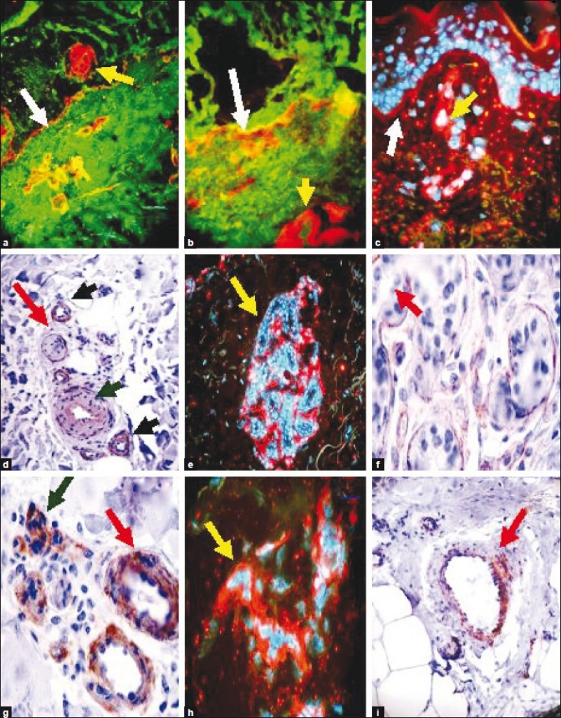
Context: Type IV collagen is a type of collagen found primarily in the skin within the basement membrane zone. The type IV collagen C4 domain at the C-terminus is not removed in post-translational processing, and the fibers are thus link head-to-head, rather than in a parallel fashion. Also, type IV collagen lacks a glycine in every third amino-acid residue necessary for the tight collagen helix. Thus, the overall collagen-IV conformation is structurally more pliable and kinked, relative to other collagen subtypes. These structural features allow collagen IV to form sheets, which is the primary structural form found in the cutaneous basal lamina. There are six human genes associated with collagen IV, specifically COL4A1, COL4A2, COL4A3, COL4A4, COL4A5 and COL4A6. The aim of this review is to highlight the significance of this protein in normal skin, and in selected diseases.
Results: The alpha 3 protein constituent of type IV collagen is thought to be the antigen implicated in Goodpasture's syndrome, wherein the immune system attacks the basement membranes of the renal glomeruli and pulmonary alveoli. In addition, mutations to the genes coding for type IV collagen lead to the Alport syndrome. Furthermore, autoantibodies directed against denatured human type IV collagen have been described in rheumatoid arthritis, scleroderma, and SLE. Structural studies of collagen IV have been utilized to differentiate between subepidermal blistering diseases, including bullous pemphigoid, acquired epidermolysis bullosa, anti-epiligrin cicatricial pemphigoid, and bullous lupus erythematosus. Collagen IV is also of importance in wound healing and in embryogenesis.
Conclusions: Pathological studies have demonstrated that minor structural differences in collagen IV can lead to distinct, clinically different diseases.
Keywords: Alport syndrome; Basement membrane zone; Collagen-type IV; Goodpasture's syndrome; Subepidermal blistering diseases.
Conflict of interest statement
Figures
References
-
- Kalluri R. Basement membranes: Structure, assembly and role in tumour angiogenesis. Nat Rev Cancer. 2003;3:422–33. - PubMed
-
- Hasegawa H, Naito I, Nakano K, Momota R, Nishida K, Taguchi T, et al. The distributions of type IV collagen alpha chains in basement membranes of human epidermis and skin appendages. Arch Histol Cytol. 2007;70:255–65. - PubMed
LinkOut - more resources
Full Text Sources
Other Literature Sources
Miscellaneous