. 2012 Mar 27;51(12):2588-96.
doi: 10.1021/bi300199d. Epub 2012 Mar 14.

Structural and biochemical characterization of the childhood cataract-associated R76S mutant of human γD-crystallin

Affiliations

Structural and biochemical characterization of the childhood cataract-associated R76S mutant of human γD-crystallin

Fangling Ji et al. Biochemistry. .

Abstract

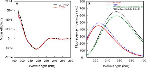
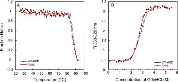
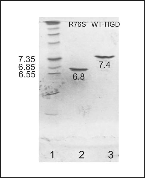
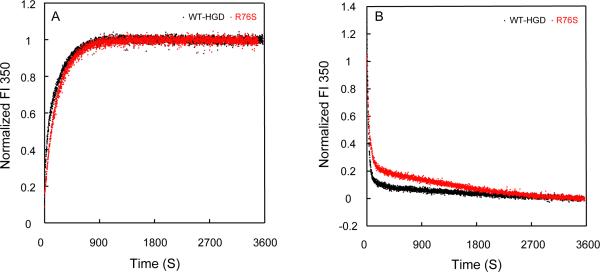
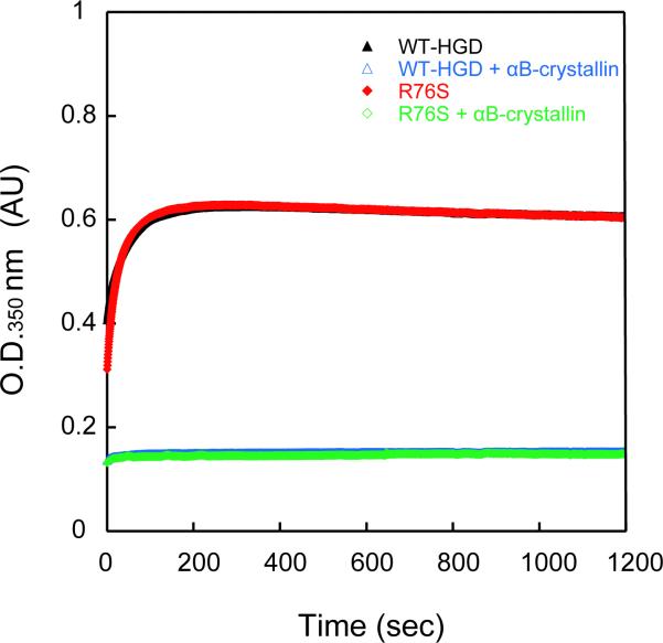
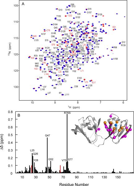
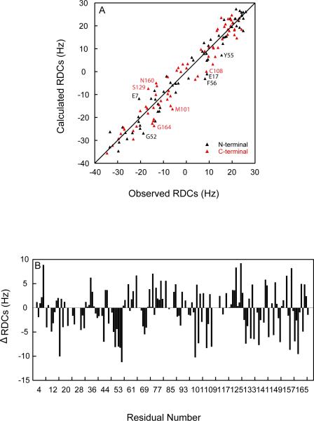
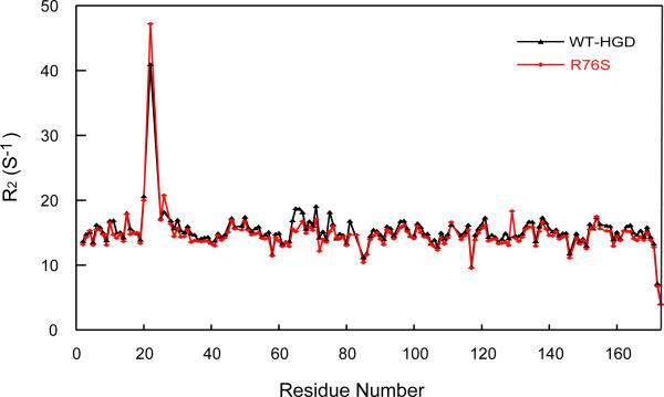
Although a number of γD-crystallin mutations are associated with cataract formation, there is not a clear understanding of the molecular mechanism(s) that lead to this protein deposition disease. As part of our ongoing studies on crystallins, we investigated the recently discovered Arg76 to Ser (R76S) mutation that is correlated with childhood cataract in an Indian family. We expressed the R76S γD-crystallin protein in E. coli, characterized it by CD, fluorescence, and NMR spectroscopy, and determined its stability with respect to thermal and chemical denaturation. Surprisingly, no significant biochemical or biophysical differences were observed between the wild-type protein and the R76S variant, except a lowered pI (6.8 compared to the wild-type value of 7.4). NMR assessment of the R76S γD-crystallin solution structure, by RDCs, and of its motional properties, by relaxation measurements, also revealed a close resemblance to wild-type crystallin. Further, kinetic unfolding/refolding experiments for R76S and wild-type protein showed similar degrees of off-pathway aggregation suppression by αB-crystallin. Overall, our results suggest that neither structural nor stability changes in the protein are responsible for the R76S γD-crystallin variant's association with cataract. However, the change in pI and the associated surface charge or the altered nature of the amino acid could influence interactions with other lens protein species.


Figures

Figure 1
Figure 1
Far-UV CD and fluorescence spectra of R76S and WT-HGD. (A) Far-UV CD spectra of R76S (dashed red line) and WT-HGD (solid black line). All samples contained 100 μg/mL protein in 10mM sodium phosphate buffer, pH 7.0 at 37 °C. (B) Fluorescence spectra of native WT-HGD (red solid line) and R76S mutant (blue dashed line) at pH7.0 and denatured WT-HGD (green dashed line) and R76S mutant (black short dotted line) in 5.5 M GdnHCl. Samples contained 10 μg/mL protein in 100 mM sodium phosphate buffer, pH 7.0, 5 mM DTT, 1mM EDTA at 37 °C.
Figure 2
Figure 2
Thermal and chemical unfolding of R76S and WT-HGD. (A) Thermal denaturation of R76S (red) and WT-HGD (black) monitored by CD spectroscopy. All samples contained 40 μg/mL purified protein in 10 mM sodium phosphate buffer, pH 7.0. (B) Chemical unfolding of R76S (red) and WT-HGD (black). All samples contained 10 μg/mL protein in 100mM sodium phosphate buffer, pH 7.0, 5 mM DTT, 1mM EDTA and GdnHCl from 0 to 5.5 M at 37 °C.
Figure 3
Figure 3
Isoelectric-focusing of R76S and WT-HGD. Marker proteins with pI values ranging from 5 to 10.5 are shown in lane 1, lanes 2 and 3 contain R76S and WT-HGD, respectively.
Figure 4
Figure 4
Kinetic unfolding/refolding experiments of R76S and WT-HGD. (A) Kinetic unfolding for R76S (red) and WT-HGD (black) at 37 °C. All samples contained 10 μg/mL protein in 100 mM sodium phosphate buffer, pH 7.0, 5 mM DTT, 1 mM EDTA. Unfolding was initiated by injecting folded native protein into a solution containing 5.5 M GdnHCl. (B) Kinetic refolding for R76S (red) and WT-HGD (black). All samples contained 10 μg/mL protein in 100 mM sodium phosphate buffer, pH 7.0, 5 mM DTT, 1mM EDTA at 37 °C. Refolding was initiated by injecting unfolded protein in 5.5 M GdnHCl into a solution containing 1.0 M GdnHCl. Fluorescence was monitored at 350 nm and the intensity was normalized with respect to native and unfolded protein controls.
Figure 5
Figure 5
Refolding of R76S and WT-HGD in the presence of the αB-crystallin chaperone. R76S and WT-HGD were unfolded in 5.0 M GdnHCl for 24 h at 37 °C. Refolding was initiated by dilution to 0.5M GdnHCl at 37 °C. The final HGD protein concentration was 50 μg/mL in all samples and the final αB-crystallin protein concentration was 250 μg/mL.
Figure 6
Figure 6
Superposition of the 1H-15N HSQC spectra and chemical shift differences between R76S and WT-HGD. (A) 1H-15N HSQC spectra of ~1 mM R76S (red contours) and WT-HGD (blue contours) at 25 °C. 162/168 amide resonances were assigned and are labeled by amino acid name and number. Residues with 1H,15N chemical shift differences > 0.05 ppm compared to WT-HGD are labeled in red. The amide resonance of R76 in WT-HGD is labeled in blue. (B) Combined amide 1H,15N chemical shift differences versus residue number. Unassigned resonances are shown with an arbitrary value of 0.1 ppm. The inset depicts the backbone structure of WT-HGD (1HK0) onto which the chemical differences are mapped. The location of R76 is marked by a blue sphere. Positions of residues whose amide resonances exhibit Δδ > 0.1 ppm and 0.1 ppm > Δδ > 0.05 ppm are shown with orange and magenta spheres, respectively.
Figure 7
Figure 7
Comparison between observed and calculated 1H,15N RDCs. (A) Calculated RDCs were predicted based on the crystal structure of HGD (PDB: 1hk0) and experimental values were measured in 5% C12E5/hexanol (r=0.96) at 25°C. Red and black data points relate to resonances of residues in the N-terminal (3–81) and C-terminal (89–171) domains, respectively. (B) Difference between observed and calculated RDC values are plotted versus residue number.
Figure 8
Figure 8
Backbone 15N R2 transverse relaxation rates for R76S (red) and WT-HGD (black). All NMR samples contained 1 mM protein in 10 mM MES buffer, pH 6.2, 5 mM DTT. All data were collected at 600 MHz and 25 °C.

References

    1. Sharma KK, Santhoshkumar P. Lens aging: effects of crystallins. Biochim Biophys Acta. 2009;1790:1095–1108. - PMC - PubMed
    1. Truscott RJ, Augusteyn RC. Oxidative changes in human lens proteins during senile nuclear cataract formation. Biochim Biophys Acta. 1977;492:43–52. - PubMed
    1. Truscott RJ, Augusteyn RC. The state of sulphydryl groups in normal and cataractous human lenses. Exp Eye Res. 1977;25:139–148. - PubMed
    1. Hains PG, Truscott RJ. Post-translational modifications in the nuclear region of young, aged, and cataract human lenses. J Proteome Res. 2007;6:3935–3943. - PubMed
    1. Ma Z, Hanson SR, Lampi KJ, David LL, Smith DL, Smith JB. Age-related changes in human lens crystallins identified by HPLC and mass spectrometry. Exp Eye Res. 1998;67:21–30. - PubMed

Publication types

MeSH terms