. 2012 May 31;379(1-2):15-22.
doi: 10.1016/j.jim.2012.02.014. Epub 2012 Feb 28.

Histamine release and surface CD200R1 staining as sensitive methods for assessing murine mast cell activation

Affiliations

Histamine release and surface CD200R1 staining as sensitive methods for assessing murine mast cell activation

David Larson et al. J Immunol Methods. .

Abstract

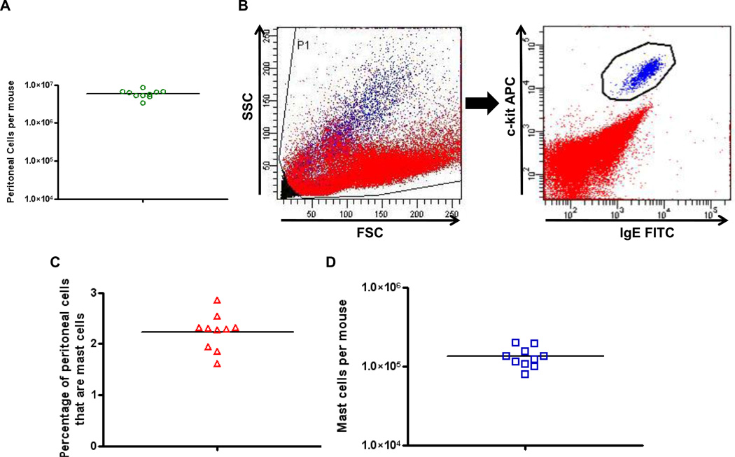
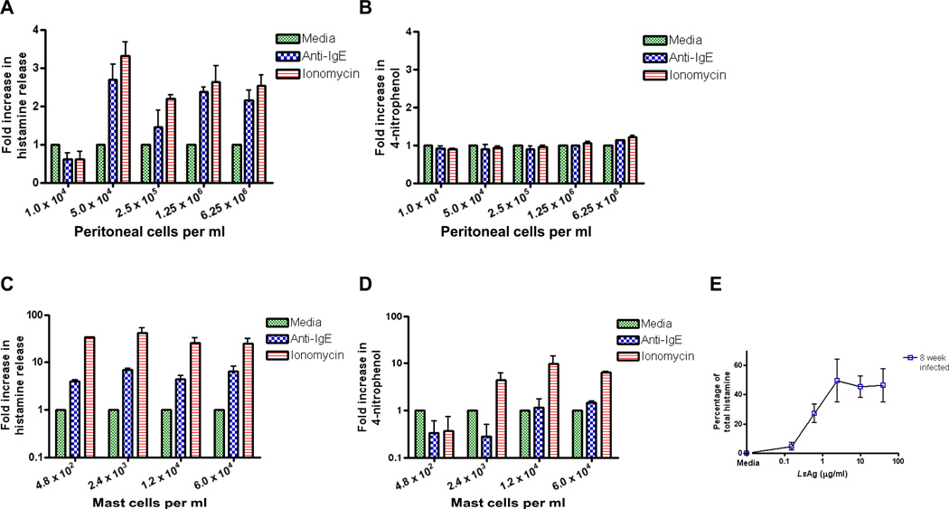
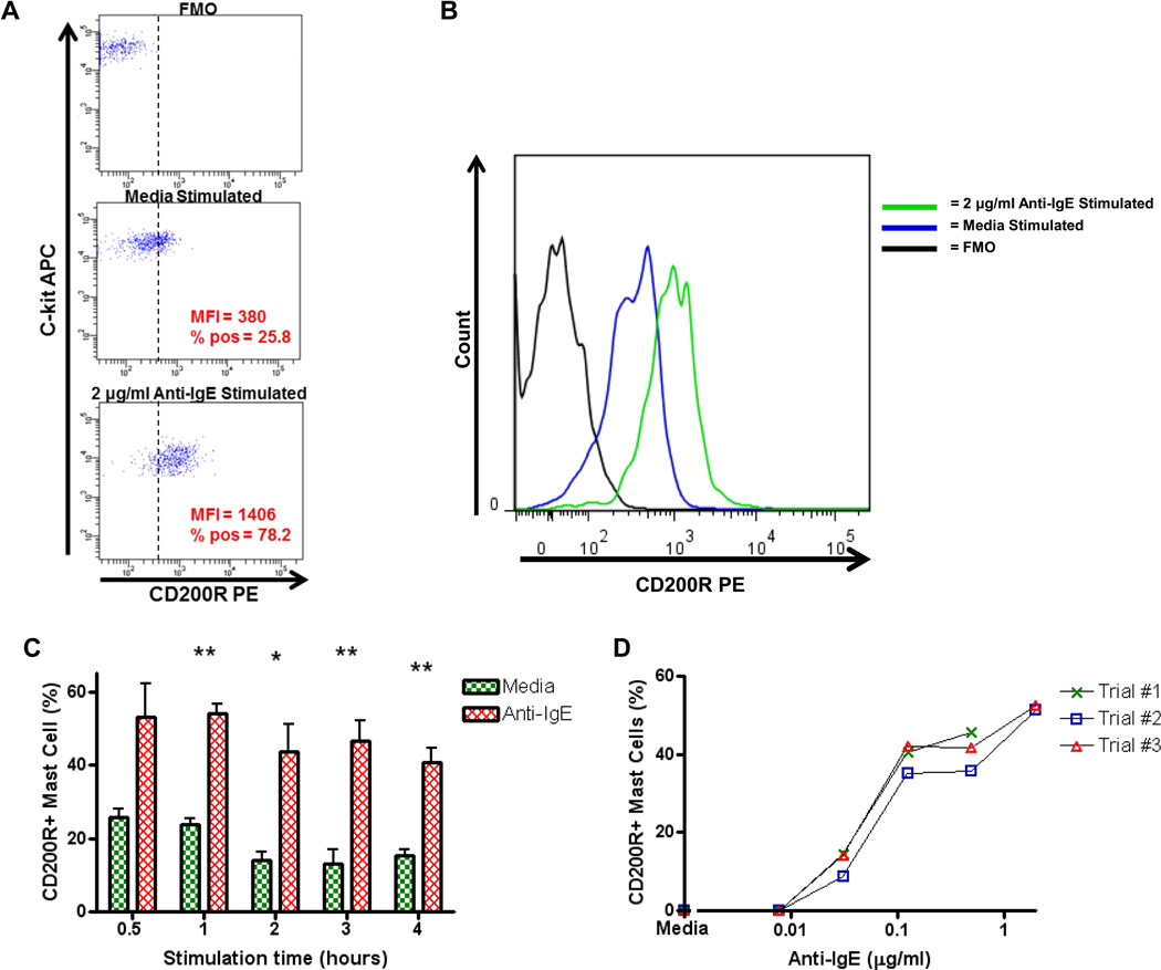
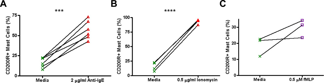
Mast cells are important effector cells of allergy and are involved in the pathology of many other diseases. Measurement of β-hexosaminidase activity, the most commonly used method for evaluation of murine mast cell activity, requires a large number of cells and thus is of limited utility for studying mast cells in mouse models of disease. In this study we evaluated the sensitivity of histamine release as compared to β-hexosaminidase activity in the measurement of mast cell activation. Whereas a minimum of 6×10(4) mast cells per ml were required to detect slight increases in β-hexosaminidase activity after anti-IgE and ionomycin stimulation, substantial increases in histamine release could be detected under the same activating conditions with as few as 480 mast cells per ml. These findings demonstrate that measurement of histamine release is substantially more sensitive than assessment of β-hexosaminidase activity for detecting mast cell activation. Additionally, we describe a novel flow cytometric method for detecting murine mast cell activation. When using 7.5×10(5) peritoneal cells per condition and gating on IgE+c-kit+cells, mast cell expression of surface CD200R1 increased after both IgE and non IgE-mediated activation. This flow cytometric procedure was uncomplicated and rapid, with increases in surface CD200R1 expression appearing after as little as 30 min of stimulation time. Measuring histamine release and surface CD200R1 expression are sensitive approaches for detection of murine mast cell activation. Further, both approaches can be done on unpurified peritoneal cell populations. By requiring low numbers of cells, these approaches are ideal for investigating mast cell activation in murine models of disease.


Figures

Figure 1
Figure 1
Identification and enumeration of peritoneal cells and peritoneal mast cells. A. Numbers of total peritoneal cells from individual mice. Peritoneal cells were obtained by lavage and counted with a hemocytometer. B. Gating strategy to identify mast cells. After initial forward and side scatter gating (left panel), mast cells were identified as IgE+ and c-kit+ cells (right panel). Mast cells shown in blue, other peritoneal cells shown in red. C. Percentages of peritoneal cells that were mast cells determined by flow cytometry. D. Total calculated number of peritoneal mast cells per mouse. For all panels, n = 10.
Figure 2
Figure 2
A 70% Percoll gradient results in a highly purified population of peritoneal mast cells. A. Cytospin of total peritoneal cells stained with May Grünwald/Giemsa stain. Mast cells determined to be 2.5% of all peritoneal cells. B. Cytospin of May Grünwald/Giemsa stained mast cells purified from the peritoneal cavity with percoll. Mast cells determined to be >98% of all cells. C. Additional staining of purified mast cells with Toluidine Blue Stain. Percentage of cells that were mast cells was identical to May Grünwald/Giemsa stainied cells. Magnification = 400x.
Figure 3
Figure 3
Histamine release is a more sensitive test of mast cell activation than β-hexosaminidase activity. (A) Histamine release and (B) β-hexosaminidase activity measured in the supernatants of peritoneal cells after 30 minute stimulation with media, 0.5 µg/ml anti-IgE, and 1 µg/ml ionomycin. (C) Histamine release and (D) β-hexosaminidase activity measured in the supernatants of purified peritoneal mast cells after 30 minute stimulation with media, 0.5 µg/ml anti-IgE, and 1 µg/ml ionomycin. E. Histamine release from 4×10³ mast cells/ml purified from the peritoneal cavity of mice infected for 8 weeks with Litomosoides sigmodontis after stimulation with LsAg. For panels A-D, fold increases in histamine release and 4-nitrophenol production were calculated by dividing anti-IgE and ionomycin stimulation levels by media stimulation levels. n = at least 3 independent experiments per condition for all experiments. Means +/− SEMs are shown.
Figure 4
Figure 4
Mast cell surface CD200R1 expression following activation with anti-IgE. A. Dot plot graphs of CD200R1 expression on peritoneal mast cells. Peritoneal cells were incubated for 3 h with media alone or 2 µg/ml of anti-IgE and then analyzed by flow cytometry. Mast cells were gated as IgE+ c-kit+ (as per figure 1B) and then evaluated for surface CD200R1 expression. Top panel shows PE staining in the absence of anti-CD200R1-PE staining (FMO, fluorescence minus one). The FMO panel was used to set a cut-off for CD200R1 positivity (dashed vertical line). MFI values are for the entire population of mast cells. % pos = % of all mast cells positive for CD200R1. B. Activation of identical mast cells shown in panel A displayed as a histogram. C. Percentages of mast cells expressing CD200R1 after stimulation of peritoneal cells with media and 0.125 µg/ml anti-IgE for 0.5, 1, 2, 3, and 4 hours (n = 3 per condition). Means +/− SEMs are shown. D. Percentages of mast cells expressing CD200R1 in response to stimulation with 0.0078, 0.031, 0.125, 0.5, and 2.0 µg/ml anti-IgE for 3 hours and after subtracting media stimulation levels. * = p < 0.05, ** = p < 0.01.
Figure 5
Figure 5
Increased surface CD200R1 expression following IgE-mediated and non IgE-mediated stimulation. Percentages of mast cells expressing surface CD200R1 after stimulation with (A) 2 µg/ml anti-IgE, (B) 0.5 µg/ml ionomycin, and (C) 0.5 µM fMLP. *** = p < 0.001, **** = p < 0.0001.

References

    1. Baumgarth N, Roederer M. A practical approach to multicolor flow cytometry for immunophenotyping. J Immunol Methods. 2000;243:77–97. - PubMed
    1. Bischoff SC. Role of mast cells in allergic and non-allergic immune responses: comparison of human and murine data. Nat Rev Immunol. 2007;7:93–104. - PubMed
    1. Cherwinski HM, Murphy CA, Joyce BL, Bigler ME, Song YS, Zurawski SM, Moshrefi MM, Gorman DM, Miller KL, Zhang S, Sedgwick JD, Phillips JH. The CD200 receptor is a novel and potent regulator of murine and human mast cell function. J Immunol. 2005;174:1348–1356. - PubMed
    1. Cohen JS, Brown HA. Phospholipases stimulate secretion in RBL mast cells. Biochemistry. 2001;40:6589–6597. - PubMed
    1. de Almeida Buranello PA, Moulin MR, Souza DA, Jamur MC, Roque-Barreira MC, Oliver C. The lectin ArtinM induces recruitment of rat mast cells from the bone marrow to the peritoneal cavity. PLoS One. 2010;5:e9776. - PMC - PubMed

Publication types

LinkOut - more resources