. 2012 Jun 14;119(24):5782-94.
doi: 10.1182/blood-2011-09-380410. Epub 2012 Mar 6.

Hypoxia promotes dissemination of multiple myeloma through acquisition of epithelial to mesenchymal transition-like features

Affiliations

Hypoxia promotes dissemination of multiple myeloma through acquisition of epithelial to mesenchymal transition-like features

Abdel Kareem Azab et al. Blood. .

Abstract

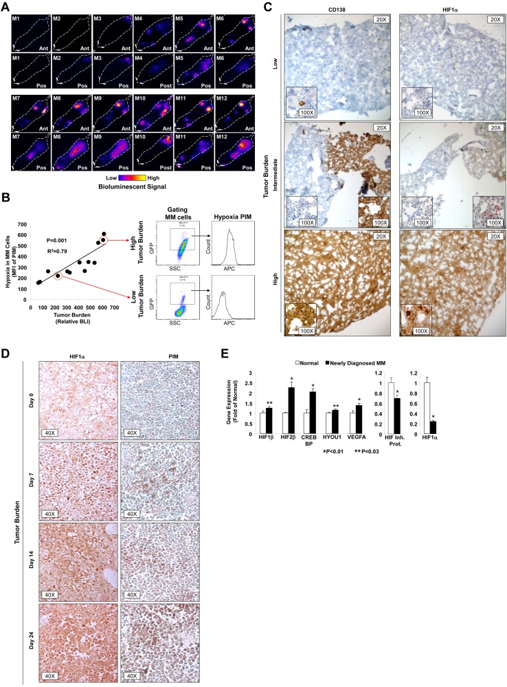
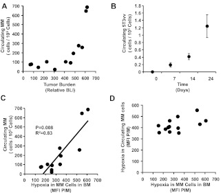
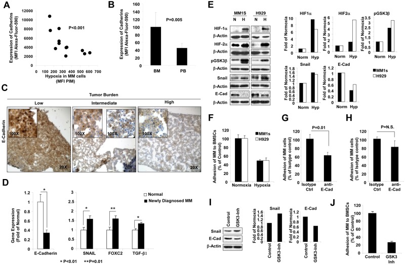
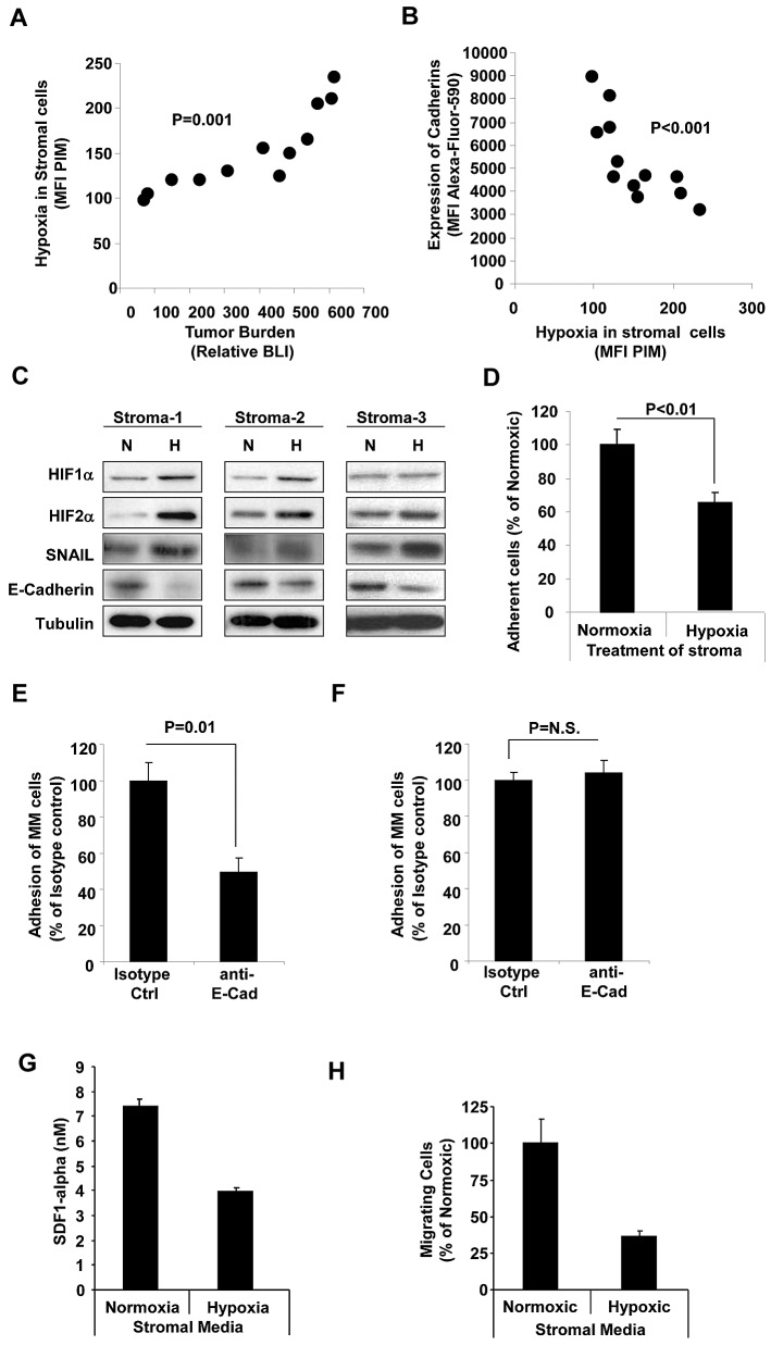
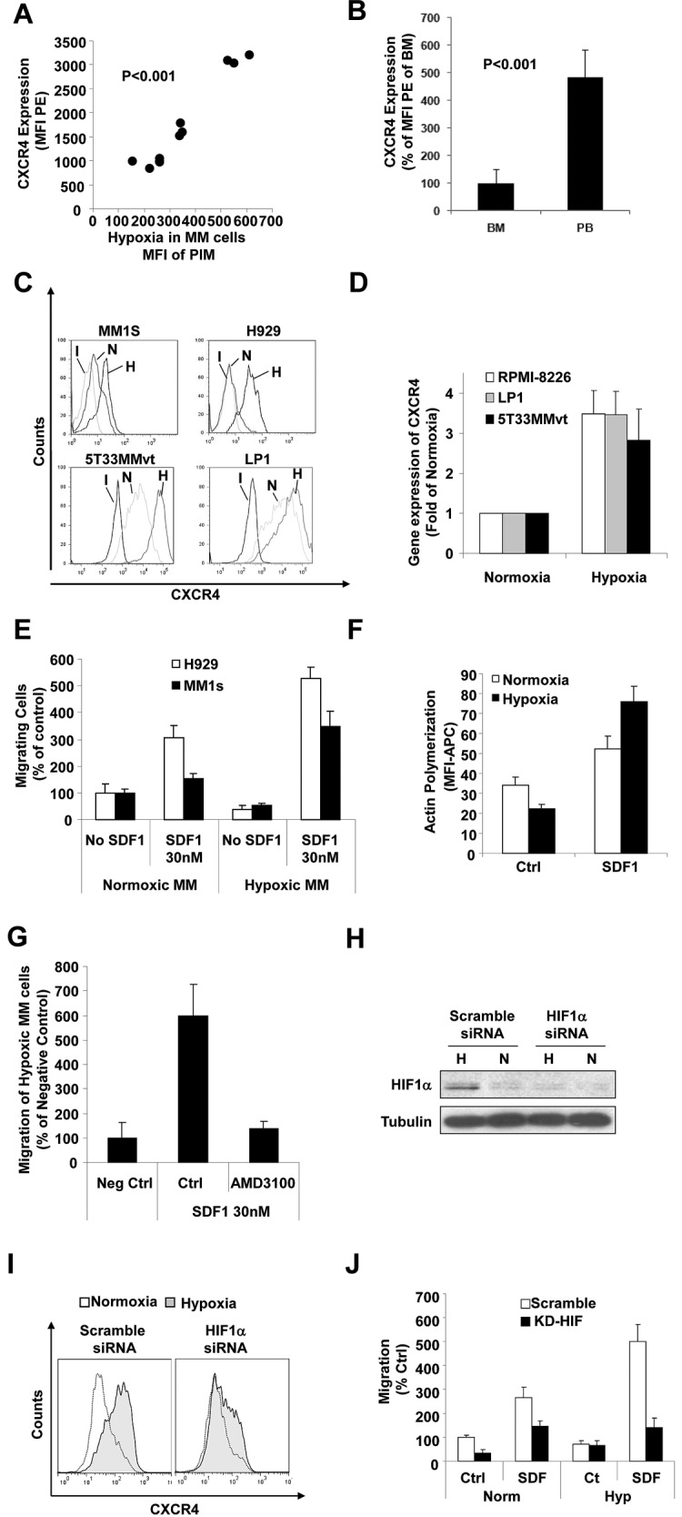
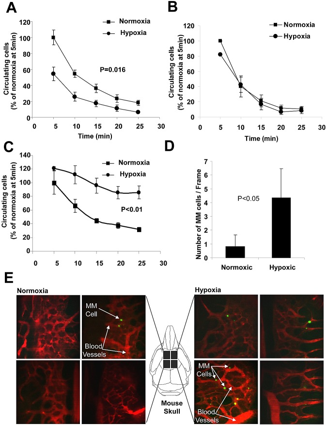
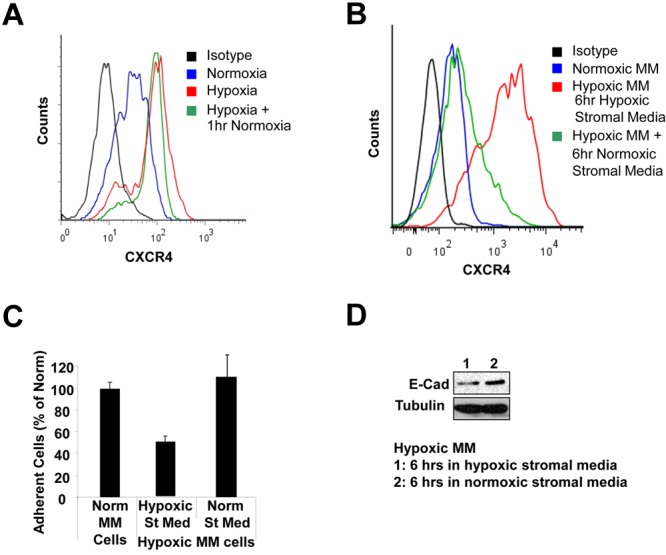
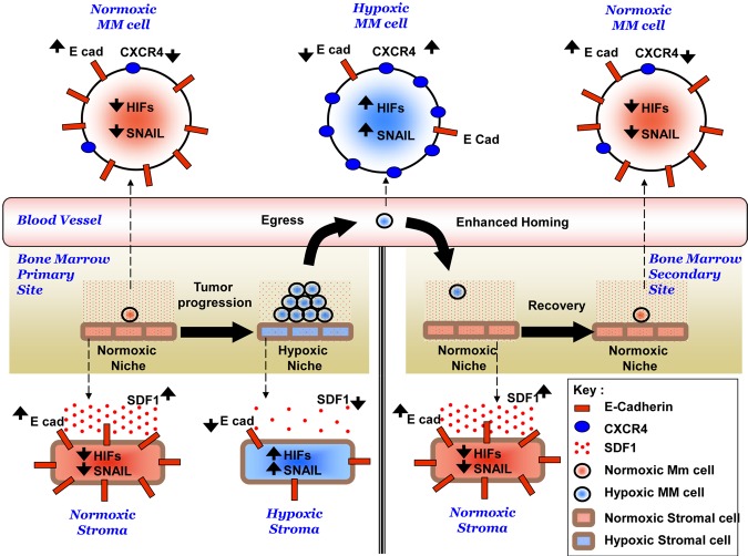
The spread of multiple myeloma (MM) involves (re)circulation into the peripheral blood and (re)entrance or homing of MM cells into new sites of the BM. Hypoxia in solid tumors was shown to promote metastasis through activation of proteins involved in the epithelial-mesenchymal transition (EMT) process. We hypothesized that MM-associated hypoxic conditions activate EMT-related proteins and promote metastasis of MM cells. In the present study, we have shown that hypoxia activates EMT-related machinery in MM cells, decreases the expression of E-cadherin, and, consequently, decreases the adhesion of MM cells to the BM and enhances egress of MM cells to the circulation. In parallel, hypoxia increased the expression of CXCR4, consequently increasing the migration and homing of circulating MM cells to new BM niches. Further studies to manipulate hypoxia to regulate tumor dissemination as a therapeutic strategy are warranted.


Figures

Figure 1
Figure 1
Tumor progression increases hypoxia in MM cells in vivo. (A) Representative BLIs of 12 SCID mice with different levels of tumor burden after injection of MM1s-GFP-Luc cells. (B) Correlation between tumor burden as detected by BLI and hypoxia in MM cells shown as the MFI of allophycocyanin-PIM in the MM cells (GFP+). (C) IHC images of specimens taken from the femurs of mice injected with MM1s-GFP-Luc with different levels of BLI stained with Abs for CD138 and HIF1α. Red arrows show nuclear staining with HIF1α. (D) IHC images of specimens taken from mice at different time points after the injection of 5T33MM mouse cells stained with Abs for PIM and HIF1α showing that binding of PIM and expression of HIF1α were directly correlated with tumor burden. (E) Gene-expression analysis of hypoxia-induced genes (including HIF1β, HIF2β, CREBBP, HYOU1, and VEGF1) in plasma cells isolated from normal subjects and MM patients using published datasets from the Gene Expression Omnibus by Chng et al (series number GSE 6477).
Figure 2
Figure 2
Correlation of hypoxia in MM cells with their egress into the circulation. (A-B) Correlation between the number of circulating MM cells detected by flow cytometry and tumor progression in SCID-MM1s model detected by BLI in the disseminated xenograft model (A) and in the 5T33MM model (B). Results show increased numbers of circulating MM cells mainly in the late stages of tumor progression. (C) Correlation between the number of circulating cells and the level of hypoxia in MM cells in the BM indicated as MFI of PIM, which shows a direct linear correlation. (D) Correlation between hypoxia levels in circulating MM cells and hypoxia in MM cells in the BM from mice with different tumor burdens, which shows that circulating MM cells were hypoxic, with no correlation with the hypoxia in the BM.
Figure 3
Figure 3
Effect of hypoxia on expression of E-cadherin and adhesion to BMSCs. (A) MM cells isolated from the BM of mice injected with MM1s cells at different tumor burdens were analyzed for the expression of cadherins by flow cytometry and compared with the hypoxic state in these cells indicated as the MFI of PIM in the MM cells. The expression of cadherins was decreased significantly with the increase in hypoxia in the MM cells. (B) The expression of E-cadherin in circulating MM cells compared with MM cells in the BM, showing that circulating MM cells maintained a low expression of E-cadherin. (C) IHC images of specimens taken from the femurs of mice with different tumor burdens stained with Ab for E-cadherin, which shows that the expression of E-cadherin decreased with tumor progression. (D) Gene-expression analysis of E-cadherin and EMT-related genes (including SNAIL, FOXC2, and TGFβ1) in plasma cells isolated from normal subjects and MM patients using published datasets from the Gene Expression Omnibus by Chng et al (series number GSE 6477). This shows decreased E-cadherin expression and increased expression of SNAIL, FOXC2, and TGFβ1 in MM. (E) The effect of incubation of MM cells (MM1s and H929) under hypoxic conditions for 24 hours in vitro on expression of HIF1α and HIF2α GSK3β, SNAIL, and E-cadherin in MM cells detected by immunoblotting. (F) The effect of incubation of MM cells (MM1s and H929) under hypoxic conditions for 24 hours in vitro on adhesion to a monolayer of BMSCs isolated from MM patients. This shows decreased adhesion of hypoxic MM cells. The effect of treating MM cells with E-cadherin–blocking Ab on adhesion of MM cells to stromal cells under normoxic (G) or hypoxic (H) conditions. This shows that the blocking Ab significantly decreased adhesion in normoxic conditions but not in hypoxic conditions. Effect of inhibition of GSK3β by a small-molecule inhibitor under normoxic conditions on expression of SNAIL and E-cadherin detected by immunoblotting (I) and on adhesion of MM cells to a monolayer of BMSCs (J). This shows increased expression of SNAIL, decreased expression of E-cadherin, and decreased adhesion to BMSCs.
Figure 4
Figure 4
Effect of tumor progression on hypoxia and expression of E-cadherin in the BM microenvironment. (A) MNCs isolated from the BM of mice injected with MM1s. Different stages were analyzed and the hypoxic state of BM microenvironment was determined by flow cytometry as the MFI of PIM in GFP cells. The correlation between hypoxia in the BM microenvironment and tumor progression (detected by BLI) demonstrates increased hypoxia in the microenvironment with tumor progression. (B) Correlation between expression of E-cadherin and level of hypoxia in BM microenvironment showing decreased expression of E-cadherin with hypoxia. (C) Effect of incubation of BMSCs (isolated from 3 different MM patients) under hypoxic conditions for 24 hours in vitro on expression of HIF1α, HIF2α, SNAIL, and E-cadherin in MM cells detected by immunoblotting. This shows increased expression of HIF1α, HIF2α, and SNAIL and decreased expression of E-cadherin in hypoxia. (D) The effect of incubation of BMSCs (isolated from 3 different MM patients) under hypoxic conditions for 24 hours in vitro on adhesion of MM cells to a monolayer of BMSCs. This shows decreased adhesion of MM cells to hypoxic BMSCs. The effect of treating stromal cells with E-cadherin–blocking Ab on adhesion of MM cells to stroma under normoxic (E) or hypoxic (F) conditions is shown. The blocking Ab decreased adhesion to normoxic but not to hypoxic stroma. (G) Effect of hypoxia on the secretion of SDF1α from MM stroma showing decreased secretion of SDF1α under hypoxic conditions. (H) The migration of MM cells to medium from normoxic and hypoxic stroma showing decreased migration of MM cells toward the medium from hypoxic stroma.
Figure 5
Figure 5
Effect of hypoxia on the expression of CXCR4 and chemotaxis of MM cells. (A) MM cells isolated from the BM of mice injected with MM1s cells at different tumor burden were analyzed for the expression of CXCR4 by flow cytometry and compared with the hypoxic state in these cells as shown by the MFI of PIM in the MM cells. This shows that the expression of CXCR4 increased significantly with the increase in hypoxia in MM cells. (B) Expression of CXCR4 in MM cells isolated from the PB and BM of mice injected with MM1s cells, which shows a higher expression of CXCR4 in circulating MM cells compared with MM cells residing in the BM. Shown is the effect of incubation of MM cells (MM1s and H929) under hypoxic conditions for 24 hours in vitro on the expression of CXCR4 in MM cells (MM1s, H929, U266, and 5T33MMvt) detected at the protein level by flow cytometry (C) or detected mRNA level by quantitative RT-PCR (D). This shows that hypoxia increased the expression of CXCR4 in MM cells. (E) Effect of incubation of MM cells (MM1s and H929) under hypoxic conditions for 24 hours in vitro on chemotaxis toward SDF1α and (F) actin polymerization showing a significant increase of chemotaxis and actin polymerization in hypoxic MM cells. (G) Effect of the CXCR4 inhibitor AMD1300 on chemotaxis of hypoxic MM cells. It can be seen that chemotaxis was abolished. (H) Knockdown of HIF1α in MM1s cells showing decreased expression of HIF1α under hypoxic conditions in cells transfected with HIF1α siRNA. (I) Effect of knockdown of HIF1α on the expression of CXCR4 induced by hypoxia. This shows that knockdown of HIF1α decreased the expression of CXCR4 induced by hypoxia. (J) The effect of knockdown of HIF1α on the increased chemotaxis induced by hypoxia. This shows that knockdown of HIF1α reversed the increase of chemotaxis in response to hypoxia.
Figure 6
Figure 6
Effect of hypoxia on MM cell homing to the BM in vivo. (A) The effect of incubation of MM1s cells under hypoxic conditions for 24 hours in vitro on homing to the BM of mice after IV injection detected by in vivo flow cytometry. This showed that hypoxia accelerated the homing of MM cells to the BM. (B) Results of homing of MM1s cells with HIF1α knockdown incubated in hypoxic or normoxic conditions for 24 hours before injection into the tail veins of mice. Hypoxia did not accelerate homing of MM cells with knockdown of HIF1α. (C) Pretreatment of hypoxic and normoxic cells with the CXCR4 inhibitor reversed their homing. MM cells were incubated under hypoxic or normoxic conditions for 24 hours and injected into mice. (D) Homing of MM cells to the BM of mouse skulls was imaged 5-7 minutes after the injection using live in vivo confocal microscopy. This shows that hypoxia increased the number of MM cells that homed to the BM, shown as the mean number of MM cells in each frame or as representative images showing the increase in the number of hypoxic cells in BM niches compared with normoxic cells (E). Green indicates MM cells; red, blood vessels.
Figure 7
Figure 7
Recovery of hypoxic MM cells in a normoxic environment. (A) Hypoxic MM cells retained a high level of CXCR4 expression after exposure to 1 hour of normoxic levels of oxygen. (B) The decreased expression of CXCR4 in hypoxic MM cells after incubation for 6 hours in medium from normoxic stroma. (C) Increased adhesion properties of hypoxic MM cells after incubation for 6 hours in medium from normoxic stroma. (D) Increased E-cadherin expression in hypoxic MM cells after incubation for 6 hours in medium from normoxic stroma.
Figure 8
Figure 8
Hypothesized mechanism of role of hypoxia in the dissemination of MM. Tumor progression induces hypoxia in the MM cells and other cells in the BM microenvironment. Hypoxia activates EMT-related machinery in MM cells and stromal cells, including activation of HIFs, activation of SNAIL, and decreased expression of E-cadherin, which leads to decreased adhesion of MM cells to the BM, decreased SDF1α secretion from stroma, and enhanced egress of MM cells to the circulation. In parallel, hypoxia increases the expression of CXCR4, and consequently increases the migration and homing of MM cells in to the BM to form metastasis to new BM niches.

Comment in

  • Metastatic myeloma?
    Shain K. Shain K. Blood. 2012 Jun 14;119(24):5612-3. doi: 10.1182/blood-2012-04-417337. Blood. 2012. PMID: 22700691 No abstract available.

References

    1. Alsayed Y, Ngo H, Runnels J, et al. Mechanisms of regulation of CXCR4/SDF-1 (CXCL12)-dependent migration and homing in multiple myeloma. Blood. 2007;109(7):2708–2717. - PMC - PubMed
    1. Lapidot T, Dar A, Kollet O. How do stem cells find their way home? Blood. 2005;106(6):1901–1910. - PubMed
    1. Damiano JS, Cress AE, Hazlehurst LA, Shtil AA, Dalton WS. Cell adhesion mediated drug resistance (CAM-DR): role of integrins and resistance to apoptosis in human myeloma cell lines. Blood. 1999;93(5):1658–1667. - PMC - PubMed
    1. Hideshima T, Chauhan D, Hayashi T, et al. The biological sequelae of stromal cell-derived factor-1alpha in multiple myeloma. Mol Cancer Ther. 2002;1(7):539–544. - PubMed
    1. Pagnucco G, Cardinale G, Gervasi F. Targeting multiple myeloma cells and their bone marrow microenvironment. Ann N Y Acad Sci. 2004;1028:390–399. - PubMed

Publication types