. 2011;15(4):113-21.
doi: 10.6091/ibj.1011.2012.

Trans-differentiation of the adipose tissue-derived stem cells into neuron-like cells expressing neurotrophins by selegiline

Affiliations

Trans-differentiation of the adipose tissue-derived stem cells into neuron-like cells expressing neurotrophins by selegiline

Alireza Abdanipour et al. Iran Biomed J. 2011.

Abstract

Background: Adult stem cells (ASC) are undifferentiated cells found throughout the body. These cells are promising tools for cell replacement therapy in neurodegenerative disease. Adipose tissue is the most abundant and accessible source of ASC. This study was conducted to evaluate effect of selegiline on differentiation of adipose-derived stem cells (ADSC) into functional neuron-like cells (NLC), and also level of the neurotrophin expression in differentiated cells.

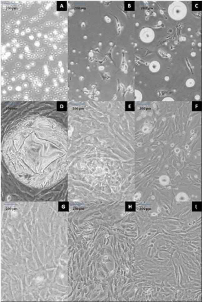
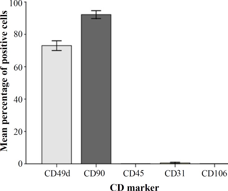
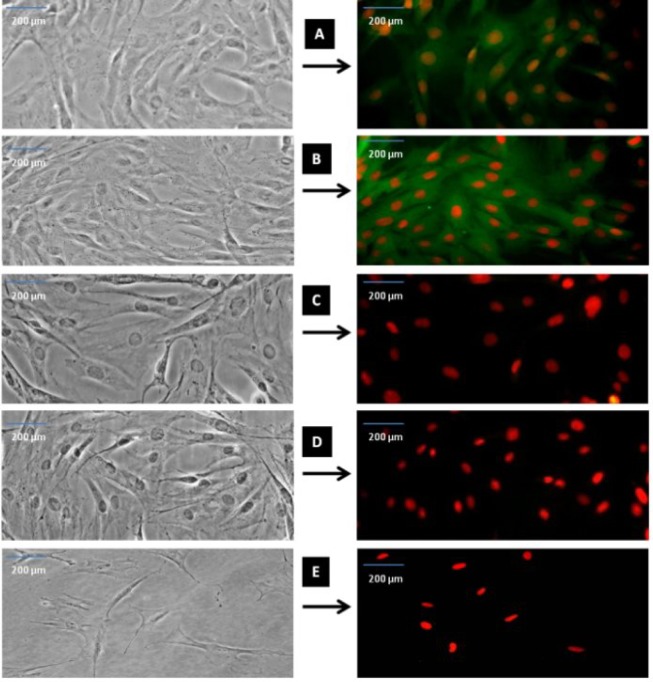
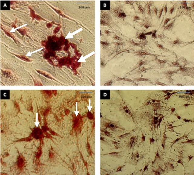
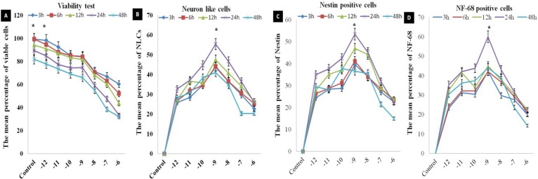
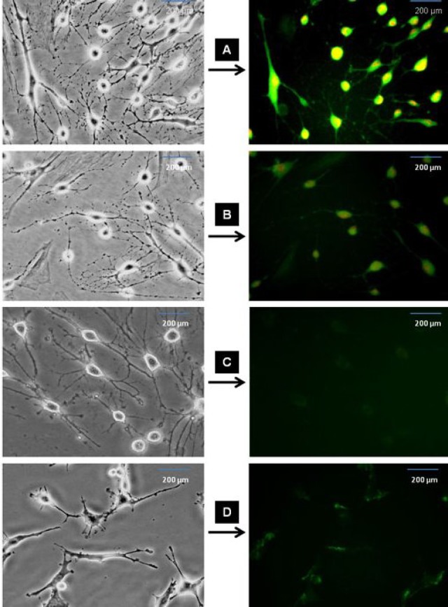
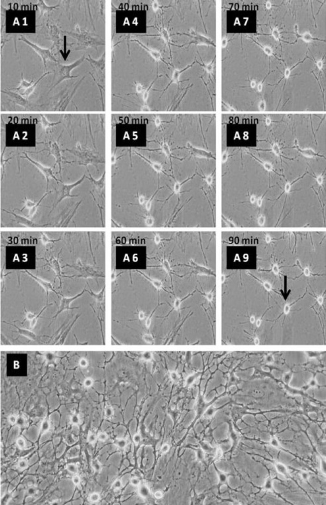
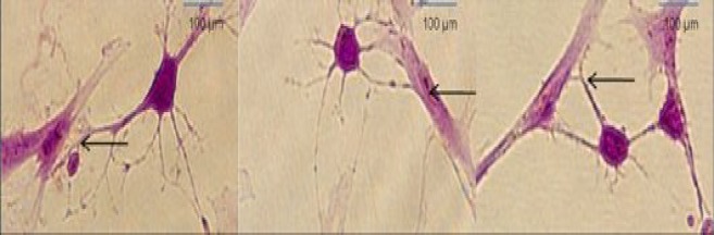
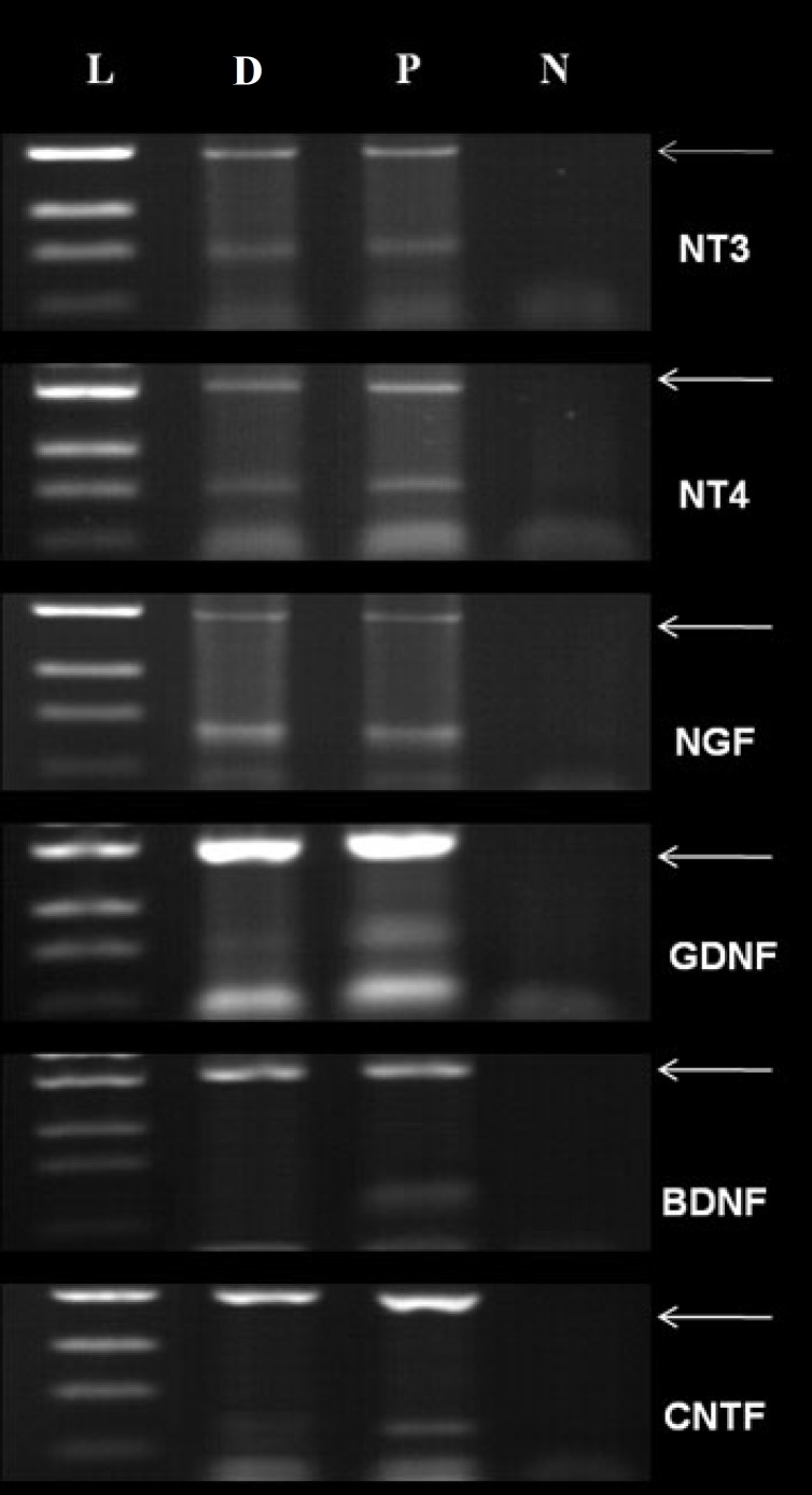
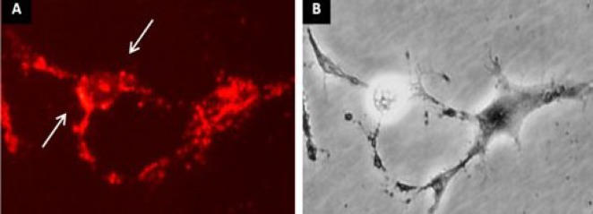
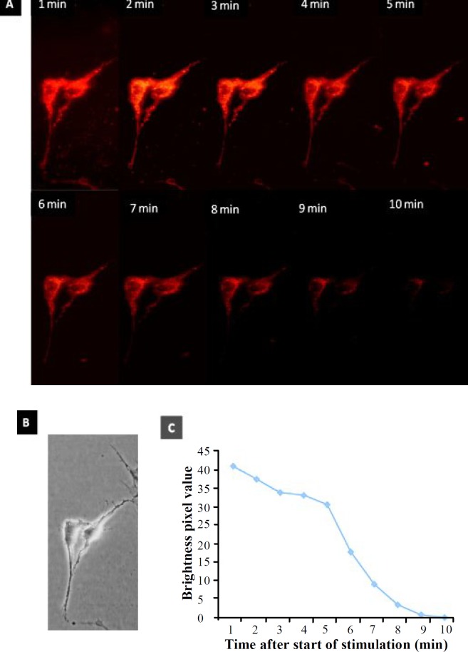
Methods: ADSC were transdifferentiated into NLC using selegiline where CD90, CD49d, CD31, CD106 and CD45 were used as markers for ADSC identification. Lipogenic and osteogenic differentiation of ADSC were used to characterize the ADSC. ADSC were treated with selegiline at different concentrations (from 10(-6) to 10(-11) mM) and time points (3, 6, 12, 24 and 48 h). Percentage of viable cells, nestin and neurofilament 68 (NF-68) immunoreactive cells were used as markers for differentiation. The optimal dose for neurotrophin expressions in differentiating cells was evaluated using reverse transcriptase-PCR. NLC function was evaluated by loading and unloading with FM1-43 dye.

Results: ADSC were immunoreactive to CD90 (95.67 ± 2.26), CD49d (71.52 ± 6.64) and CD31 (0.6 ± 0.86), but no immunoreactivity was detected for CD106 and CD45. The results of neural differentiation showed the highest percentage of nestin and NF-68 positive cells at 10(-9) mM concentration of selegiline (exposed for 24 h). The differentiated cells expressed synapsin and neurotrophin genes except brain-derived neurotrophic factor.

Conclusion: ADSC can be an alternative source in cell-based therapy for neurodegenerative diseases using selegiline to induce ADSC differentiation to neuronal lineage.


Figures

Fig. 1
Fig. 1
ADSC preparation. Floating cells at 12 h (A); attached fibroblast-like cells at (B) 24 and (C) 48 h; cultured cells with large fat droplet at day 5 (D); first (E), second (G) and third (I) passages at days 7, 11 and 13, respectively; subculture cells at days 10 (F) and 12 (H). Scale bar = 200 µm.
Fig. 2
Fig. 2
The mean percentages of immunoreactive cells to different CD markers.
Fig. 3
Fig. 3
Immunostaining of CD marker immunoreactivity after fourth passage. As representative photomicrographs shows, CD49d is specific for ADSC (A), CD90 is a specific mesenchymal stem cells (B), CD45 is a specific for hematopoietic cells (C), CD31 for endothelial cells (D) and CD106 for BMSC (E). The left sides of the fluorescent photomicrographs represent the phase contrast images from the same field of the immunofluorescence images. The cells were immunostained with relevant primary antibodies and labeled with FITC-conjugated secondary antibody (green color shows positive cells) and the red colors are ethidium bromides for counterstaining of the nuclei. Scale bar = 200 µm.
Fig. 4
Fig. 4
The in vitro osteogenesis and adipogenic differentiation. (A) ADSC after incubation for 21 days in osteogenic differentiation medium. The cells were visualized with Alizarin Red S staining. The thin arrows indicate osteoblasts and thick arrows indicate the deposition of a mineralized extracellular matrix. (B) Alizarin Red S staining of ADSC before osteogenic differentiation; (C) ADSC after incubation for 21 days in adipogenic differentiation medium. The cells were visualized with Oil Red O staining. The arrows indicate adipocytes and accumulation of fat droplets; (D) Oil Red O staining of ADSC before adipogenic differentiation. Scale bar = 100 µm.
Fig. 5
Fig. 5
The viability study in dose-response and time course evaluation of selegiline on the neuronal differentiation of the ADSC. (A) the viability of the cells exposed to various doses of selegiline and various time points and the percentage of viable cells is determined; (B) the percentage of NLC using cresyl violet staining; (C) the percentages of nestin and (D) NF-68 immunoreactive cells. These barographs show that there is a significant difference in 10-9 mM (incubated at 24 h) compared to the other time points and concentrations of selegiline (*P<0.05). The data presented as the mean ± SEM of five repeated experiments.
Fig. 6
Fig. 6
Immunofluorescence staining of differentiated ADSC into NLC using selegiline at 10-9 mM, incubated for 24 h. The cells were immunostained (left side) for the primary antibodies against nestin, NF-68, NeuN and synapsin (A, B, C and D, respectively), labeled with FITC-conjugated secondary antibody and counterstained with ethidium bromide. Right side images represent the phase contrast of the same field in the left side panel. Scale bar = 200 µm.
Fig. 8
Fig. 8
The Nissl body staining using cresyl violet. Dark blue particles in the cytoplasm show Nissl bodies and the arrows show synaptic contact regions. Scale bar = 100 µm.
Fig. 7
Fig. 7
Phase contrast images of the NLC differentiation of ADSC using selegiline. (A1-A9) time course of ADSC transdifferentiated into NLC using selegiline, incubated for 90 min. Retraction of cell body and process formation are evident at the higher time points; (B) ADSC treated with selegiline (10-9 mM, incubated for 24 h), resulted in NLC. The arrows indicate single cell prior to differentiation and the same cell after differentiation at 90 min (magnification 20×).
Fig. 9
Fig. 9
The expression of neurotrophins and GAPDH genes (internal control). L, D, P and N indicate ladder, ADSC differentiated cells into NLC, the newborn spinal cord (positive control) and negative control (without cDNA), respectively. The arrows indicate GAPDH.
Fig. 10
Fig. 10
FM 1-43 staining of ADSC differentiated into NLC. (A) The ADSC incubated with selegiline (10-9 mM) at 24 h and transdifferentiated into NLC. The arrows represent the region of fluorescent sites (synaptic vesicles) after 120 s exposure to FM1-43; (B) the phase contrast image of the same field. Scale bar = 100 µm.
Fig. 11
Fig. 11
Staining and destaining of ADSC differentiated into NLC labeled with FM1-43. (A) The fluorescence images of the FM 1-43 staining (synaptic vesicle) using 120 s exposure to FM1-43 in 10 minute after the start of the stimulation; (B) the phase contrast image of the same field; (C) total number of the pixels at the time course (1-10 min) used in the study, where the FM1-43 fluorochrome stained NLC then the cells were destained. There was a reduction in the synaptic vesicle recycling following a long time delay (magnifications 40×).

References

    1. Pachón-Peña, G , Yu, G , Tucker, A , Wu, X , Vendrell, J , Bunnell, B.A , Gimble, J.M Stromal stem cells from adipose tissue and bone marrow of age-matched female donors display distinct immune-phenotypic profiles. J. Cell Physiol. 2011;226:843–51. - PMC - PubMed
    1. Zuk, P.A , Zhu, M , Ashjian, P , Urgate, D.A , Huang, J.I , Mizuno, H , Alfonso, Z.C , Fraser, J.K , Benhaim, P , Hedrick, M.H Human adipose tissue is a source of multipotent stem cells. Mol. Biol. Cell. 2002;13:4279–4295. - PMC - PubMed
    1. Gimble, J.M , Katz, A.J , Bunnell, B.A Adipose-derived stem cells for regenerative medicine. Circ. Res. 2007;100:1249–1260. - PMC - PubMed
    1. Huang, T , He, D , Kleiner, G , Kuluz, J Neuron-like differentiation of adipose-derived stem cells from infant piglets in vitro. J. Spinal Cord Med. 2007;1:S35–40. - PMC - PubMed
    1. Zhang, D.Z , Gai, L.Y , Liu, H.W , Jin, Q.H , Huang, J.H , Zhu, X.Y Transplantation of autologous adipose-derived stem cells ameliorates cardiac function in rabbits with myocardial infarction. Chin. Med. J. (Engl). 2008;120:300–307. - PubMed

Publication types

MeSH terms