. 2012 Mar 6:3:714.
doi: 10.1038/ncomms1703.

Structure of the pentameric ligand-gated ion channel ELIC cocrystallized with its competitive antagonist acetylcholine

Affiliations
Free PMC article

Structure of the pentameric ligand-gated ion channel ELIC cocrystallized with its competitive antagonist acetylcholine

Jianjun Pan et al. Nat Commun. .
Free PMC article

Abstract

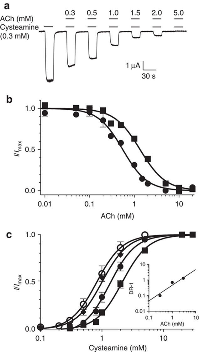
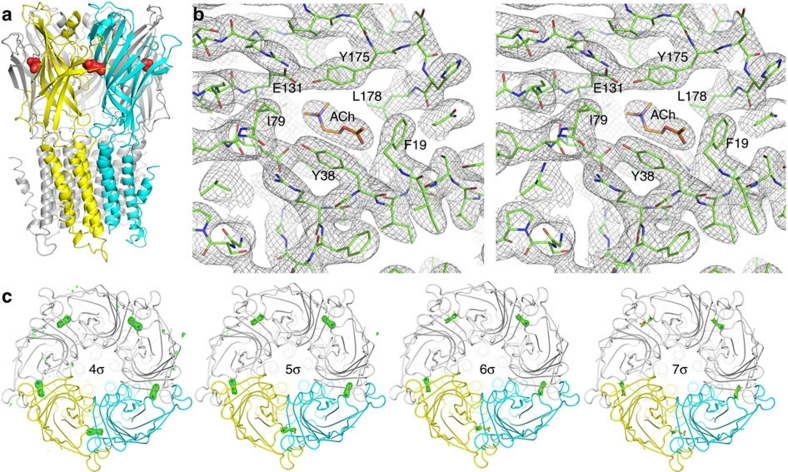
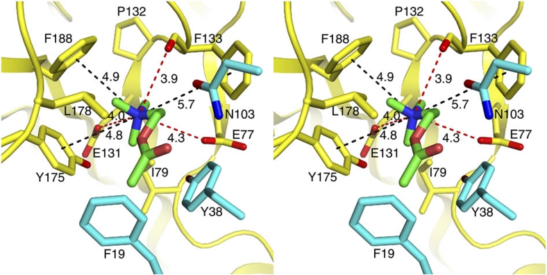
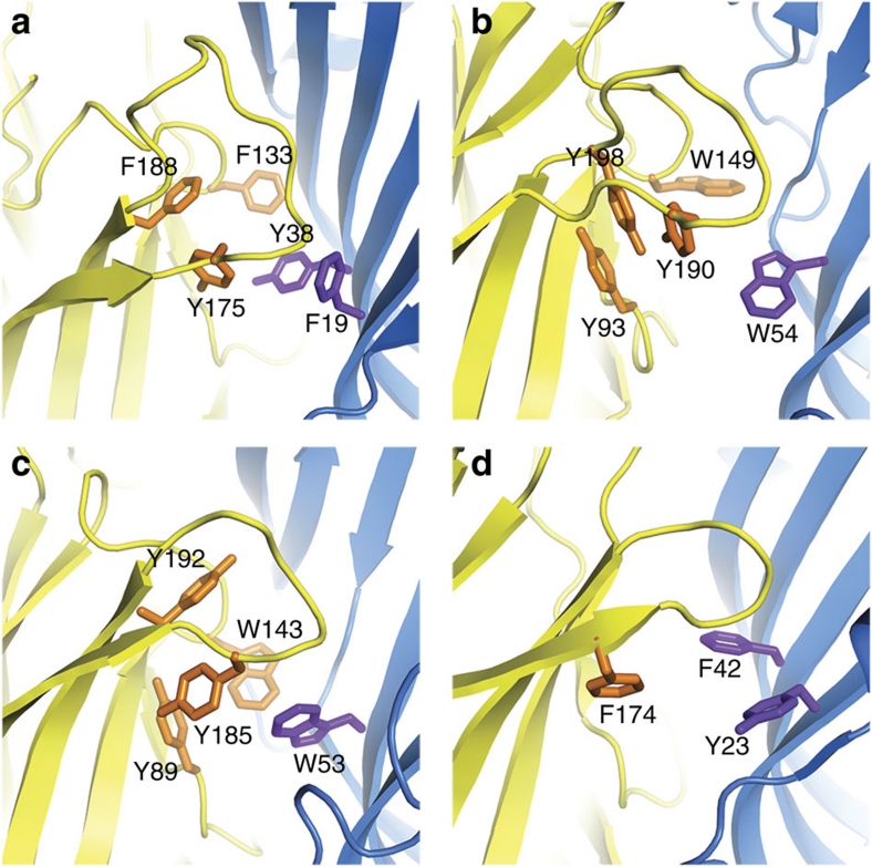
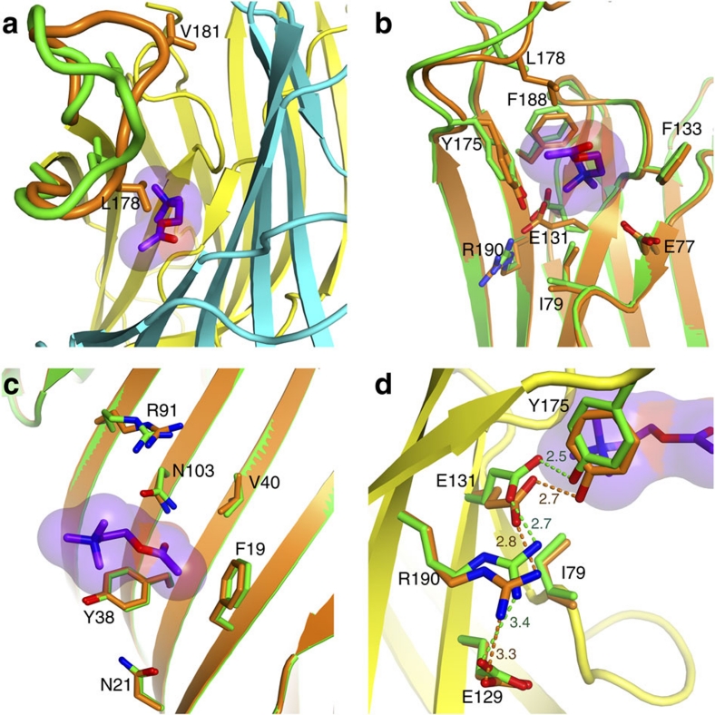
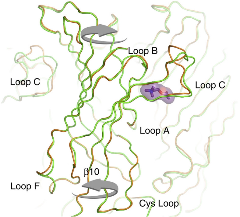
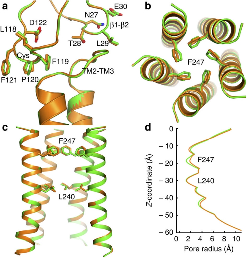
ELIC, the pentameric ligand-gated ion channel from Erwinia chrysanthemi, is a prototype for Cys-loop receptors. Here we show that acetylcholine is a competitive antagonist for ELIC. We determine the acetylcholine-ELIC cocrystal structure to a 2.9-Å resolution and find that acetylcholine binding to an aromatic cage at the subunit interface induces a significant contraction of loop C and other structural rearrangements in the extracellular domain. The side chain of the pore-lining residue F247 reorients and the pore size consequently enlarges, but the channel remains closed. We attribute the inability of acetylcholine to activate ELIC primarily to weak cation-π and electrostatic interactions in the pocket, because an acetylcholine derivative with a simple quaternary-to-tertiary ammonium substitution activates the channel. This study presents a compelling case for understanding the structural underpinning of the functional relationship between agonism and competitive antagonism in the Cys-loop receptors, providing a new framework for developing novel therapeutic drugs.


Figures

Figure 1
Figure 1. Acetylcholine inhibition of ELIC.
(a) Representative current traces in the presence of 0.3 mM cysteamine and the indicated concentrations of acetylcholine (ACh). (b) Inhibition of cysteamine-induced ELIC currents by ACh. Response is expressed as the fraction of current induced in the presence of the indicated concentrations of ACh and 0.3 mM cystamine (solid circle) or 1 mM cysteamine (solid square) relative to that in the absence of ACh. The data are fit to the Hill equation. (c) Concentration–response curves for cysteamine in the presence of 0 (open circle), 0.3 (solid diamond), 1 (solid circle) and 3 (solid square) mM ACh. The data are globally fit to a Gaddum/Schild nonlinear regression with a slope of 1. The Schild plot (inset) is also consistent with a slope of 1 (R2=0.92). All data are reported as the mean±s.e.m. from n≥7 oocytes.
Figure 2
Figure 2. The 2.9-Å-resolution structure of ELIC bound with five ACh molecules.
(a) A side view of the structure with the red surface representing ACh molecules. It is notable that the long axis of ACh is almost perpendicular to the channel axis. A pair of principal and complementary subunits for ACh binding is depicted in yellow and cyan, respectively. (b) A stereo view of the 2Fo-Fc electron density map, contoured at 1.0 σ-level, for the ACh binding site. (c) The Fo-Fc omit electron density maps contoured at different σ-levels with a carve distance of 1.8 Å for ACh molecules shown in the densities as red sticks.
Figure 3
Figure 3. Stereo view of atomic details of the ACh-binding pocket.
Dashed lines indicate distances in Å between the nitrogen atom of the positively charged quaternary ammonium of ACh and the relating residues for electrostatic interactions or potential cation-π interactions. Residues on the principal and complementary sites of the pocket are coloured in yellow and cyan, respectively. Note the position of ACh in the aromatic cage and that F133 (equivalent to W145 of the α7nACh) is likely too far away from the ACh ammonium to form a cation-π interaction.
Figure 4
Figure 4. Comparison of aromatic residues lining the ligand-binding pocket.
(a) ELIC determined in this study (PDB code: 3RQW); (b) the muscle-type nAChR between α- and γ-subunits (PDB code: 2BG9); (c) LS-AChBP (PDB code: 1UV6); and (d) GLIC (PDB code: 3EAM). The principal subunit is coloured in yellow with side chains in orange and the complementary subunit is coloured in marine with side chains in purple.
Figure 5
Figure 5. The conformational changes in and near the ACh-binding pocket.
The apo– and ACh–ELIC structures are coloured in green and orange, respectively. Structural superposition reveals conformational changes in several regions. (a) Loop C contracted substantially after ACh binding. Note that the L178 side chain is turned towards ACh. (b) Conformational changes in the principal face of the binding site. (c) Conformational changes in the principal face prompted by ACh binding are much more profound than those in the complementary face. (d) The Y175 downward movement driven by ACh binding has produced a domino effect, moving E131, R190 and E129 towards the EC–TM interface through side chain electrostatic interactions. For clarity, a and d show only the backbone structure of ACh–ELIC except for loop C in a and side chains in d, where both apo– and ACh–ELIC structures are presented. ACh is depicted in a purple surface.
Figure 6
Figure 6. 'Twist' motion in the extracellular domain on ACh binding.
Backbone superposition of the apo–ELIC (green) and the ACh–ELIC (orange) structures indicates a 'twist' motion. Several loops at the top half of the EC domain, including most visible loop C and loop B, show a counterclockwise rotation. Loop F and β10 at the bottom of the EC domain exhibit a clockwise rotation. ACh is shown in a purple surface.
Figure 7
Figure 7. Structural comparison at the EC–TM interface and the pore region.
The apo– and ACh–ELIC structures are coloured in green and orange, respectively. (a) Side chains of the residues in the β1–β2 linker (N27-E30) and the 'Cys' loop (L118-D122) are shown in stick representation. No discernable changes were observed in the β1–β2 linker. All the residues in the 'Cys' loop experienced a varying degree of side chain reorientation. (b) Top view and (c) side view of the superimposed pore region. One subunit was removed in c for clarity. No change was observed for the TM2 helical backbone. The side chain of F247 swung away from the pore centre to a small degree, yielding a less restricted pore radius near F247, as manifested by the pore radius profiles in d.

References

    1. Karlin A. Emerging structure of the nicotinic acetylcholine receptors. Nat. Rev. Neurosci. 3, 102–114 (2002). - PubMed
    1. Lester H. A., Dibas M. I., Dahan D. S., Leite J. F. & Dougherty D. A. Cys-loop receptors: new twists and turns. Trends Neurosci. 27, 329–336 (2004). - PubMed
    1. Sine S. M. & Engel A. G. Recent advances in Cys-loop receptor structure and function. Nature 440, 448–455 (2006). - PubMed
    1. Taly A., Corringer P. J., Guedin D., Lestage P. & Changeux J. P. Nicotinic receptors: allosteric transitions and therapeutic targets in the nervous system. Nat. Rev. Drug Discov. 8, 733–750 (2009). - PubMed
    1. Changeux J. P. Allosteric receptors: from electric organ to cognition. Annu. Rev. Pharmacol. Toxicol. 50, 1–38 (2010). - PubMed

Publication types

MeSH terms

Substances