

The effects of iron oxide incorporation on the chondrogenic potential of three human cell types
- PMID: 22396122
- PMCID: PMC3747461
- DOI: 10.1002/term.544
The effects of iron oxide incorporation on the chondrogenic potential of three human cell types
Abstract
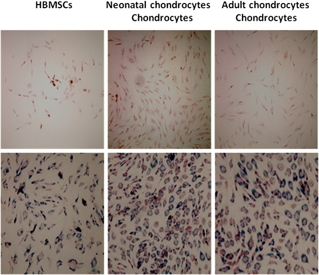
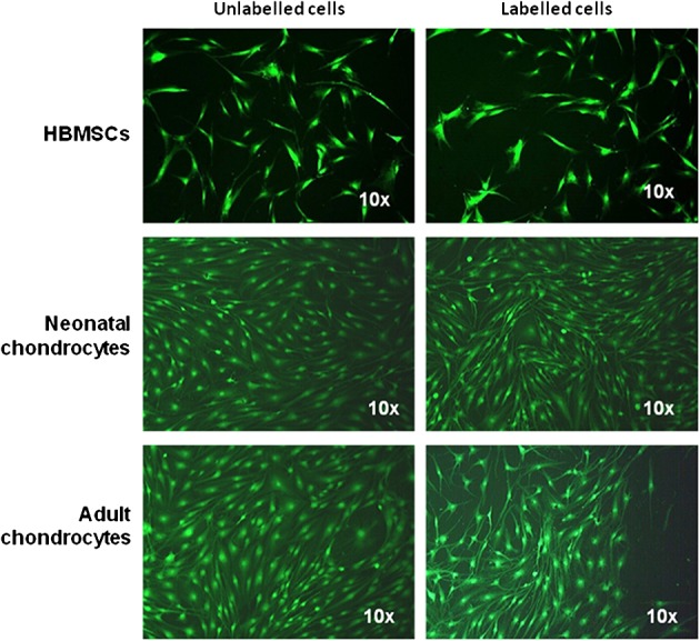
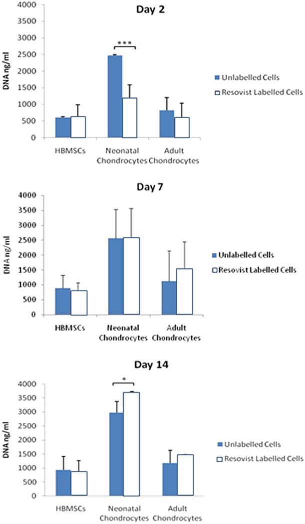
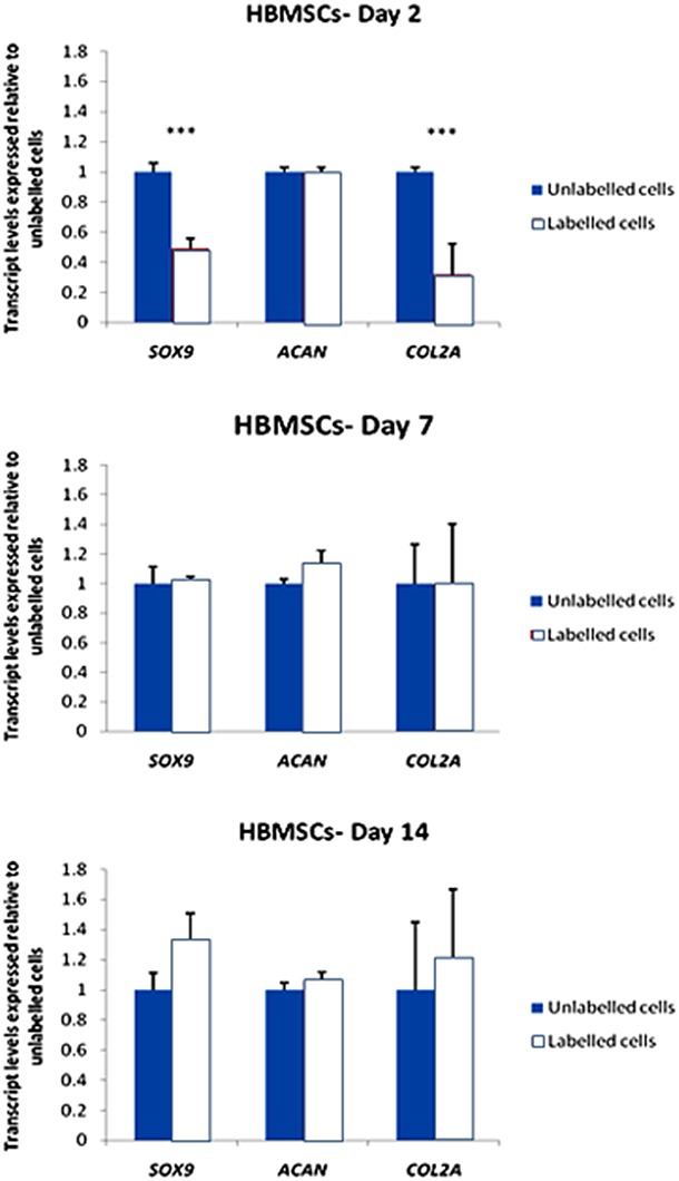
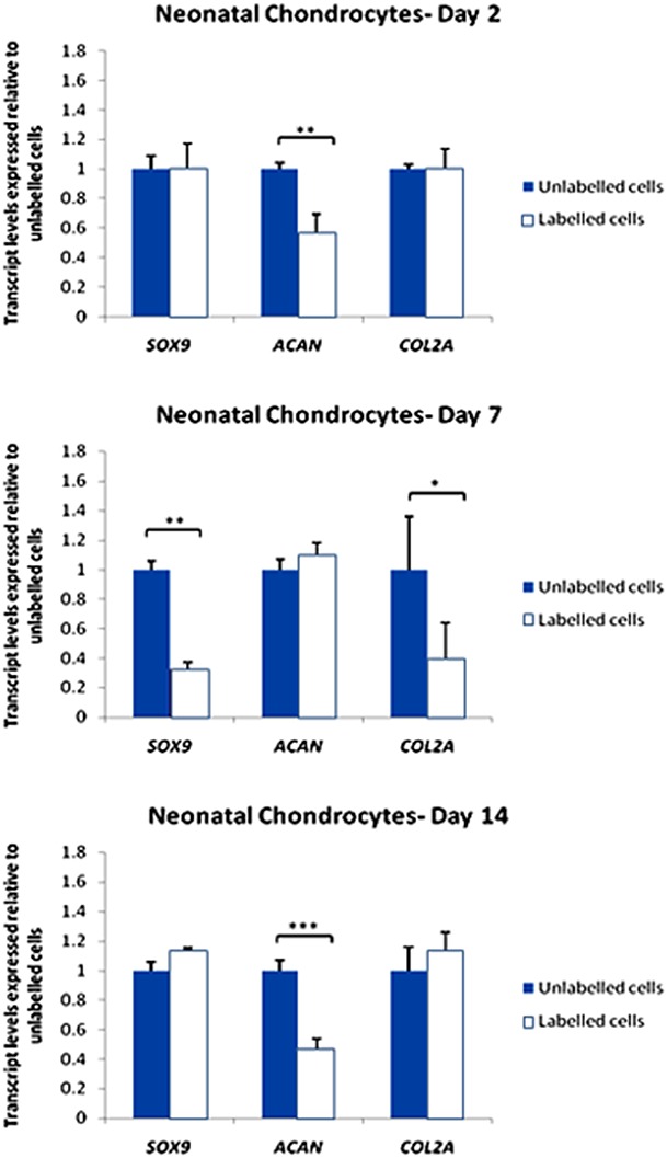
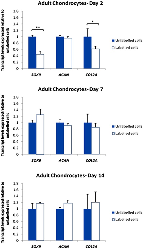
Non-invasive monitoring of living cells in vivo provides an important tool in the development of cell-based therapies in cartilage tissue engineering. High-resolution magnetic resonance imaging (MRI) has been used to monitor target cell populations in vivo. However, the side-effects on cell function of the labelling reagents, such as superparamagnetic iron oxide (SPIO), are still unclear. This study investigated the effect of SPIO particles on the chondrogenic differentiation of human bone marrow stromal cells (HBMSCs), neonatal and adult chondrocytes in vitro. Cells were labelled with SPIO for 24 h and chondrogenesis induced in serum-free medium including TGFβ3. For labelled/unlabelled cells, viability, morphology and proliferation were determined using CellTracker™ Green and PicoGreen dsDNA assays. The expression of SOX9, COL2A1 and ACAN was investigated using qRT-PCR after 2, 7 and 14 days. The results showed that viability was unaffected in all of the cells but cell morphology changed towards a 'stretched' phenotype following SPIO uptake. Cell proliferation was reduced only for labelled neonatal chondrocytes. SOX9 and COL2A1 expression decreased at day 2 but not at days 7 and 14 for labelled HBMSCs and adult chondrocytes; ACAN expression was unaffected. In contrast, SOX9 and COL2A1 expression were unaffected in labelled neonatal chondrocytes but a decrease in ACAN expression was seen at day 14. The results suggest that downregulation of chondrogenic genes associated with SPIO labelling is temporary and target cell-dependent. Resovist® can be used to label HBMSCs or mature chondrocytes for MR imaging of cells for cartilage tissue engineering.
Copyright © 2012 John Wiley & Sons, Ltd.
Figures
Similar articles
-
Dose-response of superparamagnetic iron oxide labeling on mesenchymal stem cells chondrogenic differentiation: a multi-scale in vitro study.PLoS One. 2014 May 30;9(5):e98451. doi: 10.1371/journal.pone.0098451. eCollection 2014. PLoS One. 2014. PMID: 24878844 Free PMC article.
-
Cell viability and chondrogenic differentiation capability of human mesenchymal stem cells after iron labeling with iron sucrose.Stem Cells Dev. 2014 Nov 1;23(21):2568-80. doi: 10.1089/scd.2014.0153. Epub 2014 Aug 25. Stem Cells Dev. 2014. PMID: 25036548
-
Clinically translatable cell tracking and quantification by MRI in cartilage repair using superparamagnetic iron oxides.PLoS One. 2011 Feb 23;6(2):e17001. doi: 10.1371/journal.pone.0017001. PLoS One. 2011. PMID: 21373640 Free PMC article.
-
Comparison of incremental concentrations of micron-sized superparamagnetic iron oxide for labelling articular cartilage derived chondroprogenitors.Acta Histochem. 2019 Oct;121(7):791-797. doi: 10.1016/j.acthis.2019.07.004. Epub 2019 Jul 18. Acta Histochem. 2019. PMID: 31326114
-
[Effect of superparamagnetic iron oxide on differentiation of rat bone marrow stem cells into chondrocytes in vitro].Nan Fang Yi Ke Da Xue Xue Bao. 2017 May 20;37(5):652-658. doi: 10.3969/j.issn.1673-4254.2017.05.14. Nan Fang Yi Ke Da Xue Xue Bao. 2017. PMID: 28539289 Free PMC article. Chinese.
Cited by
-
Informing future cartilage repair strategies: a comparative study of three different human cell types for cartilage tissue engineering.Cell Tissue Res. 2013 Jun;352(3):495-507. doi: 10.1007/s00441-013-1586-x. Epub 2013 Mar 12. Cell Tissue Res. 2013. PMID: 23474783 Free PMC article.
-
Stem cells as delivery vehicles for regenerative medicine-challenges and perspectives.World J Stem Cells. 2018 May 26;10(5):43-56. doi: 10.4252/wjsc.v10.i5.43. World J Stem Cells. 2018. PMID: 29849930 Free PMC article. Review.
-
The Role of Cartilage Stem/Progenitor Cells in Cartilage Repair in Osteoarthritis.Curr Stem Cell Res Ther. 2023;18(7):892-903. doi: 10.2174/1574888X17666221006113739. Curr Stem Cell Res Ther. 2023. PMID: 36201278 Free PMC article.
-
Non-invasive monitoring of in vivo hydrogel degradation and cartilage regeneration by multiparametric MR imaging.Theranostics. 2018 Jan 13;8(4):1146-1158. doi: 10.7150/thno.22514. eCollection 2018. Theranostics. 2018. PMID: 29464005 Free PMC article.
-
Dose-response of superparamagnetic iron oxide labeling on mesenchymal stem cells chondrogenic differentiation: a multi-scale in vitro study.PLoS One. 2014 May 30;9(5):e98451. doi: 10.1371/journal.pone.0098451. eCollection 2014. PLoS One. 2014. PMID: 24878844 Free PMC article.
References
-
- Afizah H, Yang Z, Hui JH, et al. A comparison between the chondrogenic potential of human bone marrow stem cells (BMSCs) and adipose-derived stem cells (ADSCs) taken from the same donors. Tissue Eng. 2007;13:659–666. - PubMed
-
- Arbab AS, Yocum GT, Kalish H, et al. Efficient magnetic cell labelling with protamine sulfate complexed to ferumoxides for cellular MRI. Blood. 2004a;104:1217–1223. - PubMed
-
- Arbab AS, Yocum GT, Kalish H, et al. Feride- protamine sulphate labelling does not alter differentiation of mesenchymal stem cells. Blood. 2004b;104:3412–3413.
-
- Bulte JW, Kraitchman DL, MacKay GT, et al. Chondrogenic differentiation of mesenchymal stem cells is inhibited after magnetic labelling with ferumoxides. Blood. 2004;104:3410–3413. - PubMed
-
- Contag CH. Molecular imaging using visible light to reveal biological changes in the brain. Neuroimaging Clin N Am. 2006;16:633–654. - PubMed
Publication types
MeSH terms
Substances
Grants and funding
LinkOut - more resources
Full Text Sources
Research Materials
