. 2012 Jun;61(6):1552-61.
doi: 10.2337/db11-0828. Epub 2012 Mar 6.

Stress augments insulin resistance and prothrombotic state: role of visceral adipose-derived monocyte chemoattractant protein-1

Affiliations

Stress augments insulin resistance and prothrombotic state: role of visceral adipose-derived monocyte chemoattractant protein-1

Yasuhiro Uchida et al. Diabetes. 2012 Jun.

Abstract

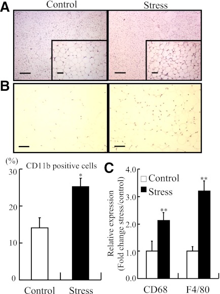
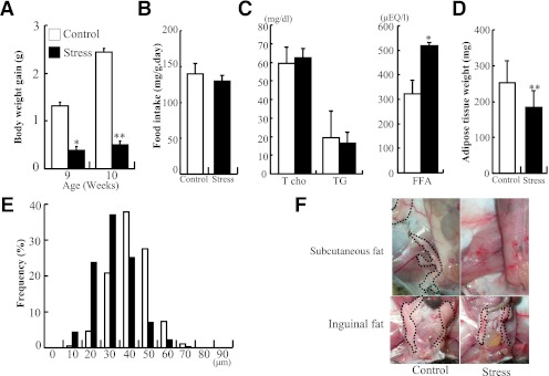
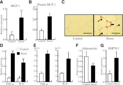
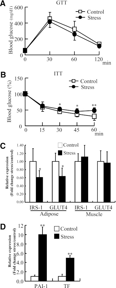
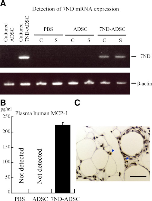
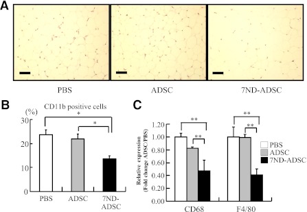
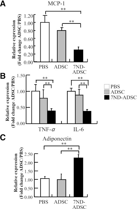
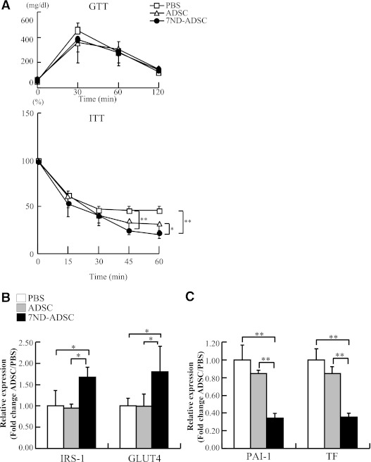
Stressors contribute to thrombosis and insulin resistance. Since obesity-related adipose inflammation is also involved in these pathological states, we assumed that stress correlates with adipose inflammation. Male mice were subjected to 2-week intermittent restraint stress. Expression of plasma lipids, monocyte/macrophage markers (CD11b, CD68, and F4/80), proinflammatory cytokines (monocyte chemoattractant protein-1 [MCP-1], tumor necrosis factor-α, and interleukin-6), adiponectin, heat shock protein 70.1 (HSP70.1), and coagulation factors (plasminogen activation inhibitor-1 [PAI-1] and tissue factor [TF]) in blood and inguinal white adipose tissue (WAT) was determined using immunohistochemistry, enzyme-linked immunosorbent assay, and RT-PCR, respectively. Glucose metabolism was assessed by glucose tolerance tests (GTTs) and insulin tolerance tests, and expression of insulin receptor substrate-1 (IRS-1) and glucose transporter 4 (GLUT4) in WAT. To examine effects of MCP-1 blockade, animals were treated with control or neutralizing antibody, or transplanted with control or 7ND (dominant-negative form of MCP-1)-overexpressing adipose-derived stromal cells (ADSCs). Stress increased monocyte accumulation, free fatty acids, proinflammatory cytokine, and HSP70.1 and reduced adiponectin. Adipose stromal cells highly expressed MCP-1. The stress-induced adipose inflammation increased PAI-1 and TF but did not give rise to thrombus formation. Without any changes in GTT, stress worsened insulin sensitivity and decreased IRS-1 and GLUT4 in WAT. Neutralizing antibody and 7ND-ADSCs reversed stress-induced adipose inflammation, procoagulant state, and insulin resistance. Stress evoked adipose inflammation to increase coagulation factors and impair insulin sensitivity through adipose-derived MCP-1.


Figures

FIG. 1.
FIG. 1.
Monocytes accumulated in inguinal adipose of stressed mice. The stressed mice were individually subjected to 2 h/day of immobilization stress for 2 weeks. Inguinal adipose tissues from stressed and control mice were analyzed by hematoxylin-eosin staining, CD11b immunostaining, and quantitative RT-PCR for CD68 and F4/80. A: Mononuclear cells accumulated in inguinal adipose tissues after the 2-week restraint stress. Original magnification ×40; bar, 250 μm (top). Original magnification × 200; bar, 50 μm (bottom). B: Accumulation of CD11b-positive cells (monocytes) increased in adipose tissue from the stressed mice (original magnification ×200; bar, 50 μm) (top). Quantitative analysis of CD11b-positive cells relative to total nuclear number (bottom). Data are expressed as mean ± SD. *P < 0.05, compared with the control mice; n = 12 in each group. C: Quantitative analysis of CD68 and F4/80 expression in adipose tissue. Data are expressed as mean ± SD. **P < 0.01, compared with the control mice; n = 12, respectively. (A high-quality digital representation of this figure is available in the online issue.)
FIG. 2.
FIG. 2.
Restraint stress reduced weight gain and adipose. Body weight and inguinal adipose of the control and stressed mice were accurately weighed, and cell sizes in the collected adipose were estimated under a microscope at ×200 magnification using image analysis software. A: Body weight gain of the control and stressed mice. B: Food intake was comparable between the control and stressed mice; n = 12 in each group. C: Plasma fat and fatty acid composition of control and stressed mice. D: Weight of inguinal adipose tissue was decreased in the stressed mice. E: Distribution of adipocyte size in inguinal adipose from the control and stressed mice. Adipose cell size was decreased in the stressed mice. F: Subcutaneous and inguinal fat pad. Circle dot lines denote adipose tissue. Data are expressed as mean ± SD. *P < 0.05 and **P < 0.01, compared with the control mice; n = 12 in each group. (A high-quality digital representation of this figure is available in the online issue.)
FIG. 3.
FIG. 3.
Restraint stress induced proinflammatory cytokine expression and reduced adiponectin expression in adipose tissue. Inguinal adipose tissues from the stressed and control mice were analyzed by quantitative RT-PCR for MCP-1, TNF-α, IL-6, and adiponectin, and by immunohistochemistry for MCP-1. Plasma level of MCP-1 from both groups was also analyzed using a mouse CCL2 ELISA kit. A: Quantitative analysis of MCP-1 expression in adipose tissue. Data are expressed as mean ± SD. **P < 0.01, compared with the control mice; n = 12 in each group. B: Plasma MCP-1 level was elevated in the stressed group. Data are expressed as mean ± SD. *P < 0.05, compared with the control mice. C: MCP-1 was highly expressed in stromal cells (arrow) and monocytes (arrowheads) in adipose tissues from the stressed mice. Original magnification ×400; bar, 25 μm. D: Quantitative analysis of TNF-α and IL-6 expression in adipose tissue. Data are expressed as mean ± SD. **P < 0.01, compared with the control mice. E: Plasma TNF-α and IL-6 levels were elevated in the stressed group. Data are expressed as mean ± SD. *P < 0.05, compared with the control mice. F: Quantitative analysis of adiponectin expression in adipose tissue. Data are expressed as mean ± SD. **P < 0.01, compared with the control mice. G: Quantitative analysis of HSP-70.1 expression in adipose tissue. Data are expressed as mean ± SD. *P < 0.05, compared with the control mice. (A high-quality digital representation of this figure is available in the online issue.)
FIG. 4.
FIG. 4.
Restraint stress worsened glucose metabolism and promoted a prothrombotic state. After 2 weeks of daily stress, intraperitoneal glucose tolerance and insulin tolerance testing were performed according to standard methods. The expressions of IRS-1, GLUT4, PAI-1, and TF in adipose tissues were analyzed with quantitative RT-PCR. A: Glucose tolerance was comparable between the control and stressed mice. B: Insulin tolerance was significantly deteriorated in the stressed mice after 30 min. Data are expressed as mean ± SD. *P < 0.05 and **P < 0.01; n = 12 in each group. C: Quantitative analysis of IRS-1 and GLUT4 expression in adipose tissue and skeletal muscle. Data are expressed as mean ± SD. *P < 0.05, compared with the control mice; n = 12, respectively. D: Quantitative analysis of PAI-1 and TF expression in adipose tissue. Data are expressed as mean ± SD. **P < 0.01, compared with the control mice.
FIG. 5.
FIG. 5.
Intraperitoneally implanted 7ND-ADSCs continuously released 7ND in vivo. PBS, ADSCs, and 7ND-ADSCs (106 cells) were injected into control and stressed mice on day 0 and 7 intraperitoneally (n = 8–10 in each group.). 7ND mRNA in inguinal adipose tissues was detected by RT-PCR using 40 cycles. Plasma level of 7ND was analyzed with human CCL2 ELISA kit. 7ND-ADSCs were detected in inguinal cells using immunohistochemistry for FLAG. A: 7ND mRNA was detected in inguinal adipose tissues from the 7ND-ADSC–treated mice but not from PBS- or ADSC-treated mice. B: 7ND protein was solely detected in peripheral blood from the 7ND-ADSC–treated mice. Data are expressed as mean ± SD. C: FLAG tag–positive cells were detected in inguinal fat of 7ND-ADSC–implanted mice. Arrowheads denote FLAG tag–positive cells. Original magnification ×400; bar, 25 μm. (A high-quality digital representation of this figure is available in the online issue.)
FIG. 6.
FIG. 6.
The 7ND-ADSC implant reduced stress-induced monocyte accumulation in the inguinal adipose. A: Mononuclear cellular accumulation was not obvious in inguinal adipose tissues from the 7ND-ADSC–treated mice (original magnification ×200 magnification; bar, 50 μm). B: Accumulation of CD11b-positive cells remarkably decreased in adipose tissue from the 7ND-ADSC–treated mice compared with control mice. Data are expressed as mean ± SD. *P < 0.05, compared with the PBS- and ADSC-treated mice, respectively; n = 8–10 in each group. C: CD68 and F4/80 expression was significantly reduced in adipose tissues from the 7ND-ADSC–treated mice. Data are expressed as mean ± SD. **P < 0.01, compared with the PBS- and ADSC-treated mice, respectively; n = 8–10 in each group. (A high-quality digital representation of this figure is available in the online issue.)
FIG. 7.
FIG. 7.
The 7ND-ADSC implant suppressed adipose inflammation in the stressed mice. A: Quantitative analysis of MCP-1 expression in adipose tissue. B: Quantitative analysis of TNF-α and IL-6 expression in adipose tissue. C: Quantitative analysis of adiponectin expression in adipose tissue. Data are expressed as mean ± SD. **P < 0.01, compared with the PBS- and ADSC-treated mice, respectively; n = 8–10 in each group.
FIG. 8.
FIG. 8.
7ND treatment rescued the stress-induced decline in insulin sensitivity and coagulability. A: Glucose tolerance was comparable among the mice treated with PBS, ADSCs, and 7ND-ADSCs (top). Insulin tolerance was significantly recovered in the 7ND-ADSC–stressed mice (bottom). Data are expressed as mean ± SD. *P < 0.05 at 60 min and **P < 0.01 at 45 and 60 min, compared with the PBS- and ADSC-treated mice, respectively; n = 8–10 in each group. B: Quantitative analysis of IRS-1 and GLUT4 expression in adipose tissue. Data are expressed as mean ± SD. *P < 0.05, compared with the PBS- and ADSC-treated mice, respectively; n = 8–10 in each group. C: Quantitative analysis of PAI-1 and TF expression in adipose tissue. Data are expressed as mean ± SD. **P < 0.01, compared with the PBS- and ADSC-treated mice, respectively; n = 8–10 in each group.

References

    1. Ziemssen T, Kern S. Psychoneuroimmunology—cross-talk between the immune and nervous systems. J Neurol 2007;254(Suppl. 2):II8–II11 - PubMed
    1. Groeschel M, Braam B. Connecting chronic and recurrent stress to vascular dysfunction: no relaxed role for the renin-angiotensin system. Am J Physiol Renal Physiol 2011;300:F1–F10 - PubMed
    1. Dimsdale JE. Psychological stress and cardiovascular disease. J Am Coll Cardiol 2008;51:1237–1246 - PMC - PubMed
    1. Kumari M, Head J, Marmot M. Prospective study of social and other risk factors for incidence of type 2 diabetes in the Whitehall II study. Arch Intern Med 2004;164:1873–1880 - PubMed
    1. Chandola T, Brunner E, Marmot M. Chronic stress at work and the metabolic syndrome: prospective study. BMJ 2006;332:521–525 - PMC - PubMed

Publication types