Review
. 2012 May;34(5):341-50.
doi: 10.1002/bies.201100098. Epub 2012 Mar 7.

Fluorescent proteins for FRET microscopy: monitoring protein interactions in living cells

Affiliations
Review

Fluorescent proteins for FRET microscopy: monitoring protein interactions in living cells

Richard N Day et al. Bioessays. 2012 May.

Erratum in

  • Bioessays. 2012 Jun;34(6):521

Abstract

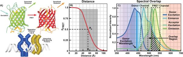
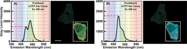
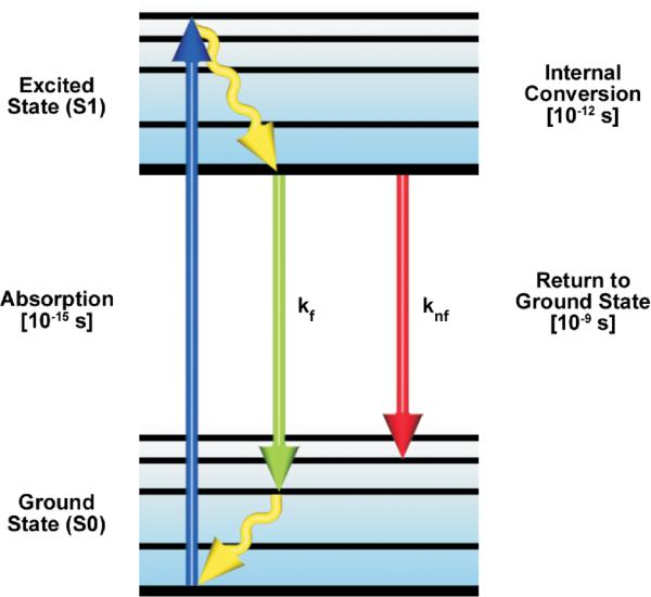
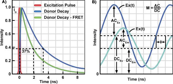
The discovery and engineering of novel fluorescent proteins (FPs) from diverse organisms is yielding fluorophores with exceptional characteristics for live-cell imaging. In particular, the development of FPs for fluorescence (or Förster) resonance energy transfer (FRET) microscopy is providing important tools for monitoring dynamic protein interactions inside living cells. The increased interest in FRET microscopy has driven the development of many different methods to measure FRET. However, the interpretation of FRET measurements is complicated by several factors including the high fluorescence background, the potential for photoconversion artifacts and the relatively low dynamic range afforded by this technique. Here, we describe the advantages and disadvantages of four methods commonly used in FRET microscopy. We then discuss the selection of FPs for the different FRET methods, identifying the most useful FP candidates for FRET microscopy. The recent success in expanding the FP color palette offers the opportunity to explore new FRET pairs.


Figures

Figure 1
Figure 1
A: Cartoon illustrating FRET between green and red FPs fused to interacting DNA-binding proteins. Energy transfer can only occur when the FPs are positioned close to one another by the interactions of the proteins they are fused to. The excitation of the green FP donor (cyan arrow) drives it to the excited-state, and that energy can be transferred directly to the nearby red FP acceptor by FRET. This results in quenching of the donor emission (green arrow) and sensitized emission (orange arrow) from the acceptor. B: The distance dependence for efficient FRET. The Förster equation (Box 1) was used to determine the change in FRET efficiency (EFRET) as a function of the separation distance between the FPs. The shaded region shows the range of 0.5 R0 to 1.5 R0 over which FRET can be accurately measured. C: The excitation and emission spectra for the Cerulean (donor) and Venus (acceptor) showing the spectral overlap between the donor emission and acceptor excitation (shaded region). The dashed boxes indicate the donor and FRET detection channels, the arrow indicates the direct acceptor excitation at the donor excitation wavelength, and the hatching shows donor SBT into the acceptor channel.
Figure 2
Figure 2
Spectral imaging of a cell producing the mTFP1-5aa-Venus fusion protein. A: The cell was illuminated at the donor excitation wavelength and spectral measurements were acquired from the ROI indicated by the red box; the calibration bar indicates 10 μm. The linked Venus fluorophore was then photobleached using the 514 nm laser line in the ROI indicated by the yellow box. This resulted in more than a 70% decrease in the Venus signal. B: The spectral measurements were then reacquired under identical conditions to the first from the same ROI (red box), and changes in the donor signal were measured. The dashed line indicates the gray level intensity of the donor signal before A) and after B) acceptor photobleaching [with permission from 59].
Figure 3
Figure 3
A simplified Perrin-Jablonski energy level diagram for a fluorescent molecule. The arrows represent absorption of excitation photon energy causing the transition from the lowest vibrational levels of the ground state (S0) to the excited state (S1). Thermal energy is lost by internal conversion and the transition from the excited state to the ground state is always from the lowest level of S1. The de-excitation transitions can occur by the emissive (kf) pathway or by other competing non-emissive (knf) pathways.
Figure 4
Figure 4
The Time Domain (TD) and Frequency-domain (FD) FLIM Methods. A: TD FLIM requires a pulsed excitation source with a femtosecond pulse width. The pulsed laser is coupled to the scanning system of the microscope. The photons emitted from the sample are recorded by a fast detector, which is connected to a time-correlated single photon counting (TCSPC) device. The TCSPC records the arrival time each photon relative to the excitation pulse, and a `photon counts' histogram is built for each pixel of an image. The fluorescence lifetime, determined as the time require for the fluorescence to decay to 37% of its initial intensity, is estimated by fitting the corresponding decay data with either single- or multi-exponential models. B: The excitation source for the FD FLIM system is a diode laser that is modulated at high radio frequencies. The emission signals from the specimen are routed to the detector, and the phase delays (Φ) and modulation ratio (M = AC/DC) of the emission (Em) relative to the excitation (Ex) are used to estimate the fluorescence lifetime.

Similar articles

Cited by

References

    1. Prasher DC, Eckenrode VK, Ward WW, Prendergast FG, et al. Primary structure of the Aequorea victoria green-fluorescent protein. Gene. 1992;111:229–33. - PubMed
    1. Chalfie M, Tu Y, Euskirchen G, Ward WW, et al. Green fluorescent protein as a marker for gene expression. Science. 1994;263:802–5. - PubMed
    1. Adams SR, Harootunian AT, Buechler YJ, Taylor SS, et al. Fluorescence ratio imaging of cyclic AMP in single cells. Nature. 1991;349:694–7. - PubMed
    1. Tsien RY. The 2009 Lindau Nobel Laureate Meeting: Roger Y. Tsien, Chemistry 2008. J Vis Exp. 2010 - PMC - PubMed
    1. Tsien RY. The green fluorescent protein. Annu Rev Biochem. 1998;67:509–44. - PubMed

Publication types

MeSH terms

Substances