

Bacteriophages φMR299-2 and φNH-4 can eliminate Pseudomonas aeruginosa in the murine lung and on cystic fibrosis lung airway cells
- PMID: 22396480
- PMCID: PMC3302570
- DOI: 10.1128/mBio.00029-12
Bacteriophages φMR299-2 and φNH-4 can eliminate Pseudomonas aeruginosa in the murine lung and on cystic fibrosis lung airway cells
Abstract
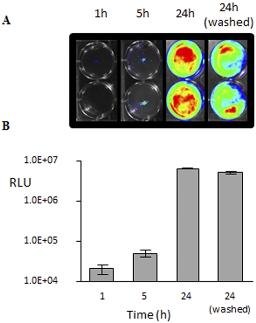
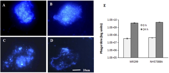
Pseudomonas aeruginosa is a common cause of infection in the lungs of patients with cystic fibrosis (CF). In addition, biofilm formation and antibiotic resistance of Pseudomonas are major problems that can complicate antibiotic therapy. We evaluated the efficacy of using bacteriophages to kill the pathogen in both biofilms and in the murine lung. We isolated and characterized two phages from a local wastewater treatment plant, a myovirus (φNH-4) and a podovirus (φMR299-2). Both phages were active against clinical isolates of P. aeruginosa. Together, the two phages killed all 9 clinical isolate strains tested, including both mucoid and nonmucoid strains. An equal mixture of the two phages was effective in killing P. aeruginosa NH57388A (mucoid) and P. aeruginosa MR299 (nonmucoid) strains when growing as a biofilm on a cystic fibrosis bronchial epithelial CFBE41o- cell line. Phage titers increased almost 100-fold over a 24-h period, confirming replication of the phage. Furthermore, the phage mix was also effective in killing the pathogen in murine lungs containing 1 × 10(7) to 2 × 10(7) P. aeruginosa. Pseudomonas was effectively cleared (reduced by a magnitude of at least 3 to 4 log units) from murine lungs in 6 h. Our study demonstrates the efficacy of these two phages in killing clinical Pseudomonas isolates in the murine lung or as a biofilm on a pulmonary cell line and supports the growing interest in using phage therapy for the control and treatment of multidrug-resistant Pseudomonas lung infections in CF patients.
Importance: Given the rise in antibiotic resistance, nonantibiotic therapies are required for the treatment of infection. This is particularly true for the treatment of Pseudomonas infection in patients with cystic fibrosis. We have identified two bacterial viruses (bacteriophages) that can kill Pseudomonas growing on human lung cells and in an animal model of lung infection. The use of bacteriophages is particularly appropriate because the killing agent can replicate on the target cell, generating fresh copies of the bacteriophage. Thus, in the presence of a target, the killing agent multiplies. By using two bacteriophages we can reduce the risk of resistant colonies developing at the site of infection. Bacteriophage therapy is an exciting field, and this study represents an important demonstration of efficacy in validated infection models.
Figures


Comment in
-
Pseudomonas biofilms, cystic fibrosis, and phage: a silver lining?mBio. 2012 Apr 3;3(2):e00061-12. doi: 10.1128/mBio.00061-12. Print 2012. mBio. 2012. PMID: 22493030 Free PMC article.
References
-
- Kerem B, et al. 1998. Identification of the cystic fibrosis gene: genetic analysis. Science 245:1073–1080 - PubMed
-
- Riordan JR, et al. 1989. Identification of the cystic fibrosis gene: cloning and characterization of complementary DNA. Science 245:1066–1073 - PubMed
-
- Rommens JM, et al. 1998. Identification of the cystic fibrosis gene: chromosome walking and jumping. Science 245:1059–1065 - PubMed
-
- Gibson RL, Burns JL, Ramsey BW. 2003. Pathophysiology and management of pulmonary infections in cystic fibrosis. Am. J. Respir. Crit. Care Med. 168:918–951 - PubMed
Publication types
MeSH terms
Substances
Associated data
- Actions
- Actions
LinkOut - more resources
Full Text Sources
Other Literature Sources
Medical
