. 2012 Apr 27;287(18):14389-401.
doi: 10.1074/jbc.M111.338434. Epub 2012 Mar 6.

Meiotic competent human germ cell-like cells derived from human embryonic stem cells induced by BMP4/WNT3A signaling and OCT4/EpCAM (epithelial cell adhesion molecule) selection

Affiliations

Meiotic competent human germ cell-like cells derived from human embryonic stem cells induced by BMP4/WNT3A signaling and OCT4/EpCAM (epithelial cell adhesion molecule) selection

Ching-Yu Chuang et al. J Biol Chem. .

Abstract

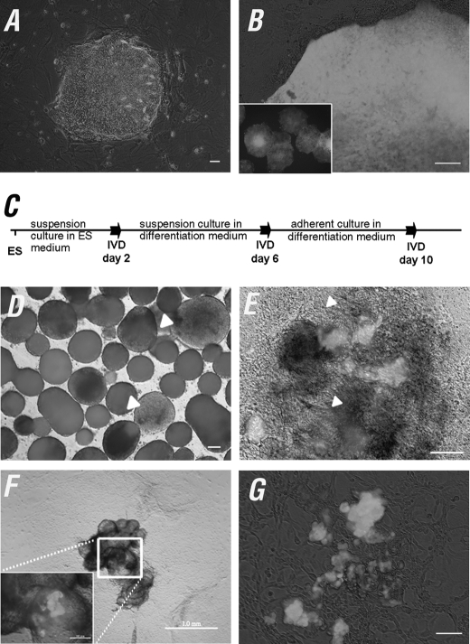
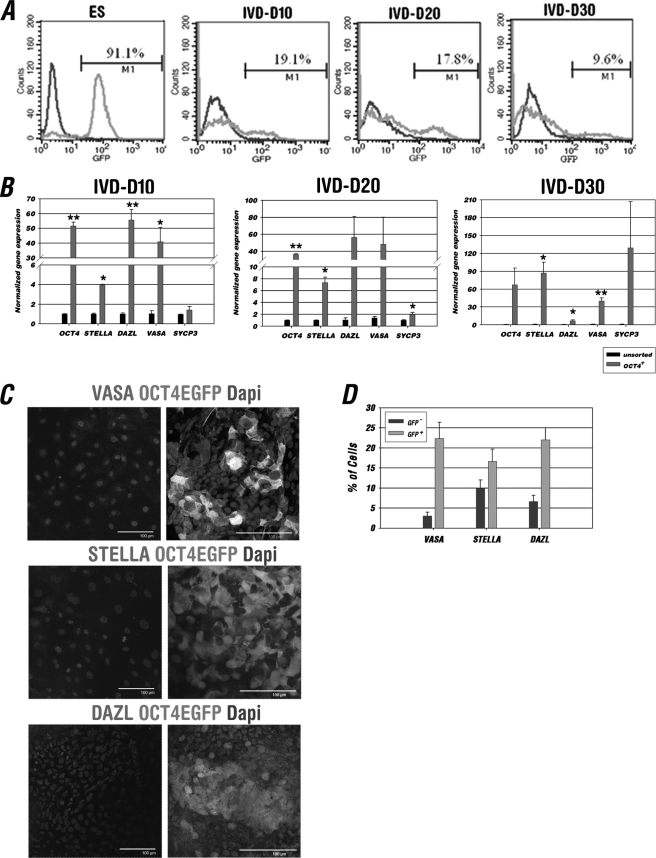
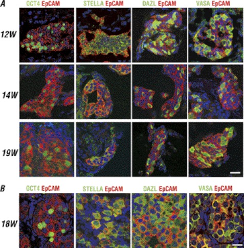
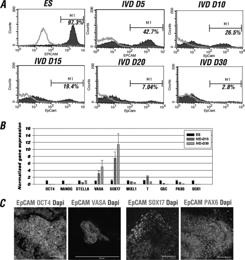
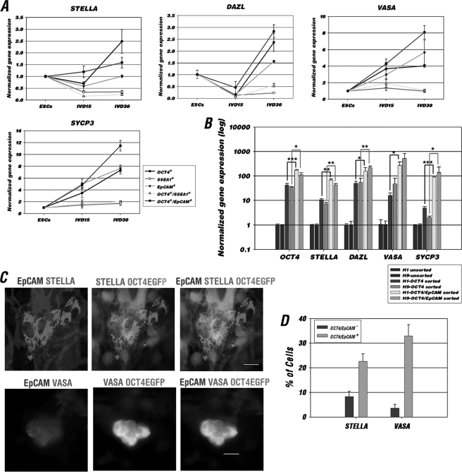
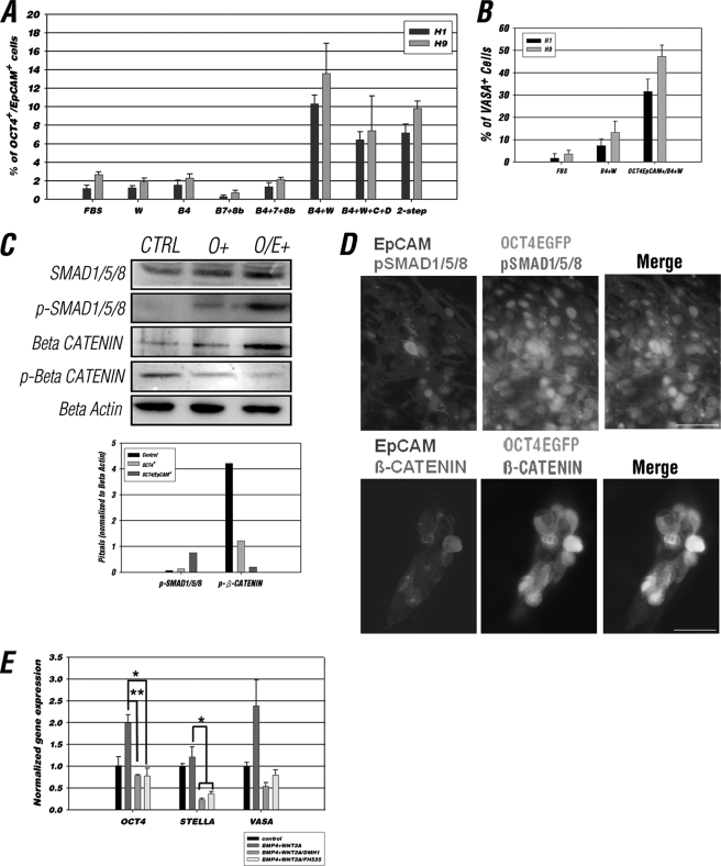
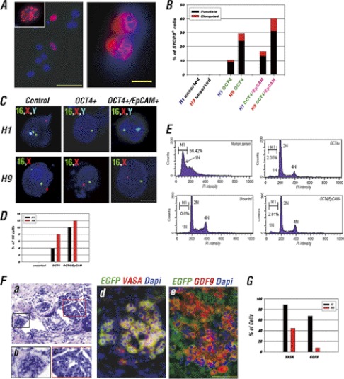
The establishment of an effective germ cell selection/enrichment platform from in vitro differentiating human embryonic stem cells (hESCs) is crucial for studying the molecular and signaling processes governing human germ cell specification and development. In this study, we developed a germ cell-enriching system that enables us to identify signaling factors involved in germ cell-fate induction from differentiating hESCs in vitro. First, we demonstrated that selection through an OCT4-EGFP reporter system can successfully increase the percentage of meiotic-competent, germ cell-like cells from spontaneously differentiating hESCs. Furthermore, we showed that the pluripotency associated surface marker, epithelial cell adhesion molecule (EpCAM), is also expressed in human fetal gonads and can be used as an effective selection marker for germ cell enrichment from differentiating hESCs. Combining OCT4 and EpCAM selection can further enrich the meiotic-competent germ cell-like cell population. Also, with the percentage of OCT4(+)/EpCAM(+) cells as readout, we demonstrated the synergistic effect of BMP4/pSMAD1/5/8 and WNT3A/β-CATENIN in promoting hESCs toward the germline fate. Combining BMP4/WNT3A induction and OCT4/EpCAM selection can significantly increase the putative germ cell population with meiotic competency. Co-transplantation of these cells with dissociated mouse neonatal ovary cells into SCID mice resulted in a homogenous germ cell cluster formation in vivo. The stepwise platform established in this study provides a useful tool to elucidate the molecular mechanisms of human germ cell development, which has implications not only for human fertility research but regenerative medicine in general.


Figures

FIGURE 1.
FIGURE 1.
OCT4-EGFP expression during hESC IVD. A and B, generation of the OCT4 promoter EGFP reporter hESC line. A, H9 hESCs were infected with lentiviruses 3 days post-colony splitting. B, the OCT4-EGFP+ cells were enriched by serial manual selection and re-plating of OCT4-EGFP+ cells until the majority (>90%) of the hESC population were OCT4-EGFP positive. C, schematic procedure for OCT4-EGFP hESCs IVD. D–G, the number of OCT4-EGFP+ cells gradually decreased during IVD. D, EBs expressed patchy EGFP during IVD 4 (white arrowhead). E, OCT4-EGFP expression was confined to clusters of cells at IVD day 10. The differentiated early neural rosettes as indicated by white arrowheads were absent from OCT4-EGFP expression. F, OCT4-EGFP+ signals were often found in the multisack structures at IVD 20 and onward. G, floating and attached OCT4-EGFP+ cell aggregates. Scale bars = 200 μm in A; 100 μm in B, D, E, and G; 1.0 mm in F, and 200 μm in the inset figure.
FIGURE 2.
FIGURE 2.
In vitro differentiated OCT4-EGFP+ cells enriched in a PGC-like population. A, FACS analysis of OCT4-EGFP hESCs and their differentiated derivatives at IVD days 10, 20, and 30. Gating was determined independently at each time point using parental H9 ES cells and their differentiated cells at each corresponding stage in parallel as a control. The average percentage of OCT4-EGFP+ cells is indicated as M1 (n = 3). B, Q-RT-PCR analysis and comparison of germline marker gene expression between the unsorted and sorted OCT4-EGFP+ cell population at IVD 10, 20, and 30. The expression value for each gene was first normalized to GAPDH and then represented as the fold-change relative to those of the unsorted group (1-fold). Data are represented as mean ± S.D. (n = 3); *, p < 0.05, **, p < 0.01. C, IF analysis of attached cell aggregates contained OCT4-EGFP (left panel) or OCT4-EGFP+ (right panel) cells at IVD 20–30 with germ cell markers VASA, STELLA, and DAZL. Little VASA, STELLA, and DAZL was detected in the cell aggregates contained EGFP cells; however, VASA, STELLA, and DAZL could be seen within the cell aggregates contained EGFP+ cells. Scale bars = 100 μm. D, quantitative analysis of positive VASA, STELLA, and DAZL IF staining in the cell aggregates contained OCT4-EGFP or OCT4-EGFP+ population. A total of 200 cells were counted in each germ cell marker staining (n = 3).
FIGURE 3.
FIGURE 3.
EpCAM co-expression with germ cell markers in human fetal testis and ovarian tissue. A, IF staining of 12-, 14-, and 19-week-old fetal testes, and B, 18-week of fetal ovarian tissue showing EpCAM was co-expressed with OCT4, STELLA, DAZL, and VASA in germ cells. Scale bars = 20 μm.
FIGURE 4.
FIGURE 4.
In vitro differentiated EpCAM-positive cells represent germ cell, and early endodermal lineages. A, FACS analysis of EpCAM positive cells during in vitro H9 hESC differentiation. Gating was determined independently at each time point using parental hESCs and their differentiated cells at each corresponding stage in parallel as a control. The average percentage of EpCAM+ cells was shown as M1 (n = 3). B, lineage-specific gene expression analysis of EpCAM-sorted H9 hESCs at IVD 15 and 30. The relative expression of each gene at IVD 15 and 30 was first normalized to GAPDH and then represented as the fold-change relative to those of the ESC group (1-fold). C, IF analysis of differentiated H9 hESCs with germ cell (OCT4 and VASA), endoderm (SOX17), and ectoderm (PAX6) markers. Scale bars = 100 μm.
FIGURE 5.
FIGURE 5.
OCT4/EpCAM selection improved germ cell-like cell enrichment. A, comparison of the expression levels of germ cell-enriched genes in OCT4-EGFP, SSEA1, EpCAM, OCT4-EGFP/SSEA1, and OCT4-EGFP/EpCAM-sorted H9 OCT4-EGFP hESCs at IVD 15 and 30. The relative expression of each gene at different IVD stages was first normalized to GAPDH and then represented as the fold-change relative to those of ESC group (1-fold). Data are represented as mean ± S.D. (n = 3). B, gene expression analysis in unsorted, OCT4-EGFP, and OCT4-EGFP/EpCAM-sorted cells at IVD 15 from H1 and H9-OCT4-EGFP hESCs. Each gene expression was first normalized to GAPDH and then represented as the fold-change relative to those of the unsorted group (1-fold) of each cell lines. Data are represented as mean ± S.D. (n = 3); ***, p < 0.001; **, p < 0.01; *, p < 0.05. C, IF staining showed co-expression of OCT4-EGFP (green) and EpCAM (blue) with germ cell markers, STELLA and VASA (red) in differentiated OCT4-EGFP hESCs. Scale bars = 20 μm. D, quantitative analysis of positive STELLA and VASA IF staining in OCT4-EGFP/EpCAM double-negative and -positive cells. A total 200 cells were counted in each staining (n = 3).
FIGURE 6.
FIGURE 6.
Combination of BMP4 and WNT3A increased the population of OCT4/EpCAM double-positive cells. A, percentage of OCT4-EGFP+/EpCAM+ cells from IVD day 15 of H1 and H9 OCT4-EGFP ESCs in 15% FBS differentiation medium or differentiation medium supplemented with various cytokines. 15% FBS differentiation medium (FBS); WNT3A (W); BMP4 (B4); BMP7 and 8b (B7 + 8b); BMP4, -7, and -8b (B4 + 7 + 8b); BMP4 and WNT3A (B4 + W); BMP4, WNT3A, SCF, and SDF (B4 + W + C + D); two-step, cells were cultured in differentiation medium supplemented with BMP4 and WNT3A for 4 days, then switched to medium with BMP4, WNT3A, SCF, and SDF. Data are presented as mean ± S.D., n = 3. B, percentage of VASA-positive cells from IVD day 15 of H1 and H9 OCT4-EGFP ESCs in cells cultured with FBS, BMP4 + WNT3A, and BMP4 + WNT3A-treated OCT4-EGFP/EpCAM double-positive cells. A total of 200 cells were counted in each staining (n = 3). C, Western blot analysis of SMAD-1/5/8, and phospho-SMAD-1/5/8, β-CATENIN, phospho-β-CATENIN in control (unpurified), OCT4-EGFP, and OCT4-EGFP/EpCAM-positive sorted cells (upper panel). Semi-quantification of the relative levels of phospho-SMAD-1/5/8 and phospho-β-CATENIN (corrected to the total levels of respective proteins) are shown in the lower panel. An equal amount of protein (20 μg) was loaded in each lane. The intensity of bands was normalized to that of β-ACTIN. D, IF analysis showed co-expression of OCT4-EGFP (green), EpCAM (blue), and phospho-SMAD1/5/8 or β-CATENIN (red) in differentiating OCT4-EGFP hESCs at IVD 15. Scale bars = 50 μm. E, comparing the gene expression level for cells differentiated in control (15% FBS medium contained vehicle (dimethyl sulfoxide)), BMP4 + WNT3A, DMH1, and FH535 added medium. The relative expression of each gene was normalized to the control group after normalization with GAPDH. Data are represented as mean ± S.D., n = 3; *, p < 0.05; **, p < 0.01.
FIGURE 7.
FIGURE 7.
OCT4/EpCAM selection in combination with BMP4 and WNT3A induction improves meiotic progression, haploid formation, and development of germ cell-like structure in kidney capsule transplantation. A, evidence of meiosis from the BMP4/WNT3A-induced in vitro differentiated germ cell-like cells as demonstrated by IF analysis with SYCP3 antibody. Meiotic nuclei with punctate (left panel) and elongated (right panel) SYCP3 staining patterns represent early and late meiosis processes, respectively. Nuclei were stained with DAPI (blue). Scale bars = 50 (left panel) and 10 μm (right panel). B, quantitative analysis of the percentage of cells with punctate or elongated SYCP3-positive signals in the unpurified, OCT4-sorted or OCT4/EpCAM-sorted cells from A. A total of 150 meiotic spread cells each were counted for H1 and H9 cell lines. C, FISH analysis on unpurified, OCT4, and OCT4/EpCAM-sorted cells at IVD day 20 of H1 and H9 OCT4-EGFP ESCs in differentiated medium containing BMP4/WNT3A with DNA probes specifically targeting chromosomes 16, X, and Y. Nuclei were stained with DAPI II (blue). Scale bars = 5 μm. D, quantitative analysis of the percentage of haploid cells with single chromosomes 16, and X or Y signal in C. A total of 100 fixed cells were counted. E, DNA content analysis of unsorted, OCT4-sorted, and OCT4/EpCAM-sorted cells in BMP4/WNT3A-induced H1 OCT4-EGFP cells. Human semen was used as a positive control to determine the gating of haploid cells (1N) by flow cytometer. The percentage of 1N cells was indicated as M1. F, characterization of hESC H9-derived OCT4+/EpCAM+ germ cells following co-transplantation with dissociated mouse fetal ovarian cells into mouse kidney capsules. a–c, H&E stain of a OCT4+/EpCAM+ cell transplanted kidney. Compact cell clusters exhibit “germ cell cluster-like” structures within the mouse kidney (b and c). d and e, IF staining of VASA (red), GDF9 (red), and EGFP (green) on cryosections of the transplanted kidney showed a uniform distribution of VASA/OCT4-EGFP or GDF9/OCT4-EGFP co-expressing cells in the germ cell-like cluster. Scale bars = 50 μm. G, quantitative comparison of the percentage of VASA+ and GDF9+ cells in the kidney capsule transplantation or IVD (day 30–40) of OCT4+/EpCAM+ H9 cells treated with BMP4/WNT3A. A total of 200 OCT4+/EpCAM+ cells were counted in the IVD group and 400 OCT4-EGFP-positive cells were counted in the kidney capsule transplantation group.

References

    1. Hübner K., Fuhrmann G., Christenson L. K., Kehler J., Reinbold R., De La Fuente R., Wood J., Strauss J. F., 3rd, Boiani M., Schöler H. R. (2003) Derivation of oocytes from mouse embryonic stem cells. Science 300, 1251–1256 - PubMed
    1. Nicholas C. R., Haston K. M., Grewall A. K., Longacre T. A., Reijo Pera R. A. (2009) Transplantation directs oocyte maturation from embryonic stem cells and provides a therapeutic strategy for female infertility. Hum. Mol. Genet. 18, 4376–4389 - PMC - PubMed
    1. Nayernia K., Nolte J., Michelmann H. W., Lee J. H., Rathsack K., Drusenheimer N., Dev A., Wulf G., Ehrmann I. E., Elliott D. J., Okpanyi V., Zechner U., Haaf T., Meinhardt A., Engel W. (2006) In vitro differentiated embryonic stem cells give rise to male gametes that can generate offspring mice. Dev. Cell 11, 125–132 - PubMed
    1. Geijsen N., Horoschak M., Kim K., Gribnau J., Eggan K., Daley G. Q. (2004) Derivation of embryonic germ cells and male gametes from embryonic stem cells. Nature 427, 148–154 - PubMed
    1. Toyooka Y., Tsunekawa N., Akasu R., Noce T. (2003) Embryonic stem cells can form germ cells in vitro. Proc. Natl. Acad. Sci. U.S.A. 100, 11457–11462 - PMC - PubMed

Publication types

MeSH terms