. 2012 Mar 20;109(12):4473-8.
doi: 10.1073/pnas.1101003109. Epub 2012 Mar 6.

Striated organelle, a cytoskeletal structure positioned to modulate hair-cell transduction

Affiliations

Striated organelle, a cytoskeletal structure positioned to modulate hair-cell transduction

Florin Vranceanu et al. Proc Natl Acad Sci U S A. .

Abstract

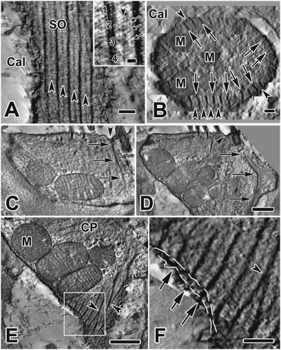
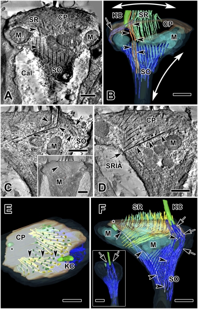
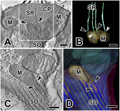
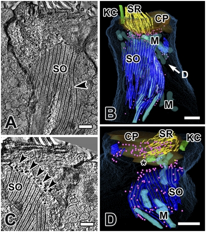
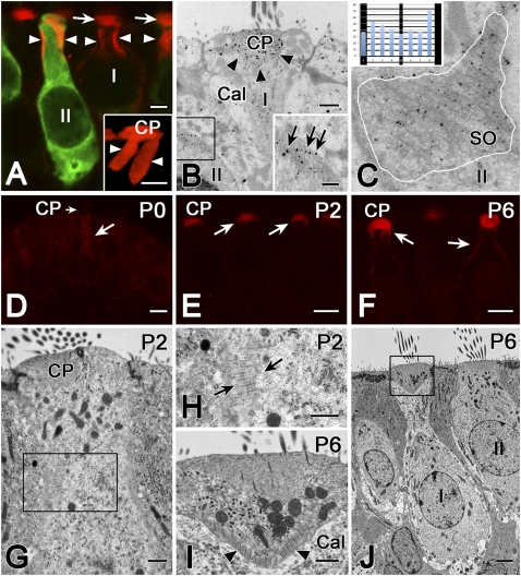
The striated organelle (SO), a cytoskeletal structure located in the apical region of cochlear and vestibular hair cells, consists of alternating, cross-linked, thick and thin filamentous bundles. In the vestibular periphery, the SO is well developed in both type I and type II hair cells. We studied the 3D structure of the SO with intermediate-voltage electron microscopy and electron microscope tomography. We also used antibodies to α-2 spectrin, one protein component, to trace development of the SO in vestibular hair cells over the first postnatal week. In type I cells, the SO forms an inverted open-ended cone attached to the cell membrane along both its upper and lower circumferences and separated from the cuticular plate by a dense cluster of exceptionally large mitochondria. In addition to contacts with the membrane and adjacent mitochondria, the SO is connected both directly and indirectly, via microtubules, to some stereociliary rootlets. The overall architecture of the apical region in type I hair cells--a striated structure restricting a cluster of large mitochondria between its filaments, the cuticular plate, and plasma membrane--suggests that the SO might serve two functions: to maintain hair-cell shape and to alter transduction by changing the geometry and mechanical properties of hair bundles.


Conflict of interest statement

The authors declare no conflict of interest.

Figures

Fig. 1.
Fig. 1.
Structure of the SO. (A) Tangential section from a type I hair cell EM tomogram showing the structure, periodicity, and location of the SO immediately subjacent to the calyx membrane. (Inset) Thick (numbered) and thin (arrowhead) filament bundles are composed of several thinner, spiral-bundled filaments (e.g., 1–4), measuring ∼10 nm and ∼6 nm, respectively. Note also the cross filaments (small arrows), which EM immunogold studies indicate are likely spectrin (Fig. 5 B and C, and Insets). (Scale bar, 0.2 μm.) (B) Cross-section through the neck of a type I cell showing radial distribution of thick (arrows) and thin (arrowheads) SO filament bundles. They extend from the hair cell membrane to a depth of ∼130 nm into the cell. (Scale bar, 0.2 μm.) (C and D) Apical insertion of a thick SO filament bundle (arrows) adjacent to the kinocilium. The site of insertion is a dimpled area (arrowhead) between the kinocilium and nearby SRs. (D) A SO filament bundle angles downward (arrows), approaching the cell membrane and continuing into the neck of the type I cell. (Scale bar, 0.5 μm; also applies to C.) (E and F) Details of hair-cell neck region in cell 2, obtained with SLICER mode in IMOD. (E) Thick filament bundles of the SO merge together (arrowheads). (Scale bar, 1 μm.) (F) Higher magnification of the area enclosed in the box in E shows possible connections (arrows) between thick filament bundles of the SO and the calyx membrane bridging the intercellular cleft (dashed white lines show locations of hair-cell and calyx membranes). (Scale bar, 0.25 μm.). Cal, calyx; CP, cuticular plate; M, mitochondria.
Fig. 2.
Fig. 2.
Tomograms and reconstructions of two type I hair cells. (A) Tomogram section of cell 1, a juxtastriolar type I cell, showing the connection between a SR (arrowhead) and a SO thick filament bundle (arrow). (B) Three-dimensional reconstruction of the same type I hair cell from a volume obtained by joining six serial tomograms (3.0 μm total thickness). The apical part of the SO extends from the lateral aspect of the cell membrane (arrow) to the apical hair cell membrane near the kinocilium. Two SRs are continuous with the SO (orange, arrowheads). SO thick (dark blue) and thin (light blue) filament bundles envelop a group of large mitochondria (transparent light blue). One possible effect on cell shape and hair bundle tension, based on filament direction and cell membrane insertions is illustrated: radial contraction of the SO elongates the neck of the hair cell (vertical two-headed arrow), which simultaneously splays the hair bundle through the connection of the rootlets with the SO (horizontal two-headed arrow). (C–F) Cell 2, an extrastriolar type I cell, modeled by joining 11 serial tomograms (total volume thickness = 5.5 μm). (C) One section from this tomogram shows a 110° bend in two of the longest rootlets (arrowheads). An apical extension of the SO (arrows) near the kinocilium (not seen in this section), is also shown in the model (see below, F and F Inset, arrows). (Inset) Section from cell 5, a striolar type I hair cell, fortuitously illustrating four bent rootlets in a single plane. (D) The lower of two SRs in C (arrowheads) traverses the cuticular plate and inserts into the SR insertion area (arrow) on the cell membrane on the opposite side of the cell. (E) A view from the top of the hair cell in C and D. Inside the cuticular plate, long, bent SRs (yellow, arrowheads), originating near the kinocilium and identical to those in C and D, traverse the cuticular plate and insert in the plasma membrane; thinner, green stereocilia (located farther from the kinocilium) do not traverse the cuticular plate. (F) Lateral view of the 3D model of cell 2. Thick (dark blue) and thin (light blue) SO filament bundles are distinguished. Arrows point to an extension of the SO that encircles the kinocilium. Nearby, a kinociliar rootlet (gray, arrowheads) extends from the kinocilium down toward the neck of the hair cell; another (arrowheads) is observed extending from the centriole (asterisk). (Inset) Viewed from the kinociliar side, the same model shows apical insertions of the SO (arrows) on each side of the kinocilium. (Scale bars, 0.5 μm in A–D, and C Inset); 1 μm in E and F.) Cal, calyx; CP, cuticular plate; KC, kinocilium; M, mitochondria.
Fig. 3.
Fig. 3.
Connections between subcuticular mitochondria and SRs or SO filaments. (A) Cell 1: SRs can terminate on subcuticular mitochondria (arrowheads); left arrowhead points to a mitochondrion that receives two distinct rootlets. (B) Model view of area enclosed in white box in A shows contacts between the distal end of several rootlets and subcuticular mitochondria. Two black arrowheads are identical to those in A; two white arrowheads point to additional mitochondrial-rootlet connections. (C and D) Cell 2: SO filaments are closely associated (arrowheads) with subcuticular mitochondria, as seen in a tomogram section (C) and in a model view (D) that highlights linkages (red) between the SO and subcuticular mitochondria. (Scale bars, 0.25 μm.) CP, cuticular plate; M, mitochondria.
Fig. 4.
Fig. 4.
The striated organelle in cell 4, an extrastriolar type II hair cell, modeled by joining nine serial tomograms (total volume thickness = 4.5 μm). (A) A large portion of the SO is visualized in a single plane in SLICER mode, illustrating the planar nature of the SO in type II cells. Morphing of a thick bundle into a thin one can be observed (arrowhead). (B) A partial 3D reconstruction of the same hair cell. “D” in B refers to the view in D (i.e., from under the cuticular plate). (C) In another section from the same hair cell, note the dense spherical objects (arrowheads, depicted as pink spheres in B and D) aligned with many of the thin filaments along the margins of the SO. (D) View from below the cuticular plate: SRs are not connected to the SO, but may be associated with the dense spherical objects (pink spheres). Most mitochondria, except for those in close association with the SO, have been removed for clarity. (Scale bars, 0.5 μm.) CP, cuticular plate; KC, kinocilium; M, mitochondria; *, centriole.
Fig. 5.
Fig. 5.
The α-2 spectrin is one protein component of the SO. (A) Confocal microscopy shows cuticular plate (arrows) and SO (arrowheads) immunoreactivity for α-2 spectrin (red). Calretinin antibody (green) distinguishes type II (II) from type I (I) hair cells. (Inset) Higher magnification Volocity reconstruction of α-2 spectrin label (red) in a type II hair cell shows the SO, which appears to hang down in two large flaps (arrowheads) from the cuticular plate (CP). (Scale bars, 2 μm.) (B) EM immunogold with an antibody to α-2 spectrin (gold particles) localizes the protein to the cuticular plate (arrowheads) in a type I cell (I) and to the SO (black box) in a type II cell (II). (Inset) Higher magnification of the area enclosed within the box in B. Gold particles (arrows) had a tendency to be located over the cross-links between the thick and thin filament bundles. (Scale bars, 0.5 μm.) (C) Quantification of the α-2 spectrin EM immunogold results. In 14 SO profiles, such as this example (SO) from a type II hair cell (II), gold particles were identified within an area delineated by a white line circumscribing the SO profile. Starting with each thick filament bundle, intervals between thick bundles were divided into eight equally spaced samples running parallel to the thick bundles, and the interval into which each particle fell was determined. (Inset) For 363 particles, sums in the eight sample bins are illustrated, superimposed upon a schematic of the SO. A χ2 test of homogeneity (χ2 = 23.1, df = 7, P < 0.002) indicated a preference for intervals immediately adjacent to the thick filament bundles. (D–F) Confocal microscopy of α-2 spectrin antibody in developing rat crista in the first postnatal week. (D) At birth (P0), there is no label in the cuticular plate (CP, short arrow), and only weak immunoreactivity in the upper half of the hair cell. (E) At P2, α-2 spectrin antibody labels the cuticular plate less intensely than at P6 (F), when it also labels parts of the lateral membrane in the region of the SO. (Scale bars, 5 μm.) (G–J) Normal TEM of developing rat hair cells. At P2 (G and H), the SO is just beginning to form in a hair cell of indeterminate type. The calyx ending, which defines type I hair cells, typically does not begin to form until P4 (21). (Scale bars in G and H, 0.5 μm.) At P6 (I and J), both the calyx (Cal) surrounding the type I cell and the SO (arrowheads) are well developed. Several stereociliar rootlets can also be observed within the cuticular plate in I. (Scale bars, 0.5 μm in I and 2 μm in J.)

References

    1. Dierkes K, Lindner B, Jülicher F. Enhancement of sensitivity gain and frequency tuning by coupling of active hair bundles. Proc Natl Acad Sci USA. 2008;105:18669–18674. - PMC - PubMed
    1. Fettiplace R, Hackney CM. The sensory and motor roles of auditory hair cells. Nat Rev Neurosci. 2006;7:19–29. - PubMed
    1. Hudspeth AJ. How the ear's works work: Mechanoelectrical transduction and amplification by hair cells. C R Biol. 2005;328:155–162. - PubMed
    1. DeRosier DJ, Tilney LG. The structure of the cuticular plate, an in vivo actin gel. J Cell Biol. 1989;109:2853–2867. - PMC - PubMed
    1. Forge A, Wright T. The molecular architecture of the inner ear. Br Med Bull. 2002;63:5–24. - PubMed

Publication types