. 2012;8(3):e1002498.
doi: 10.1371/journal.pgen.1002498. Epub 2012 Mar 1.

Insulin/IGF-1 and hypoxia signaling act in concert to regulate iron homeostasis in Caenorhabditis elegans

Affiliations

Insulin/IGF-1 and hypoxia signaling act in concert to regulate iron homeostasis in Caenorhabditis elegans

Daniel Ackerman et al. PLoS Genet. 2012.

Abstract

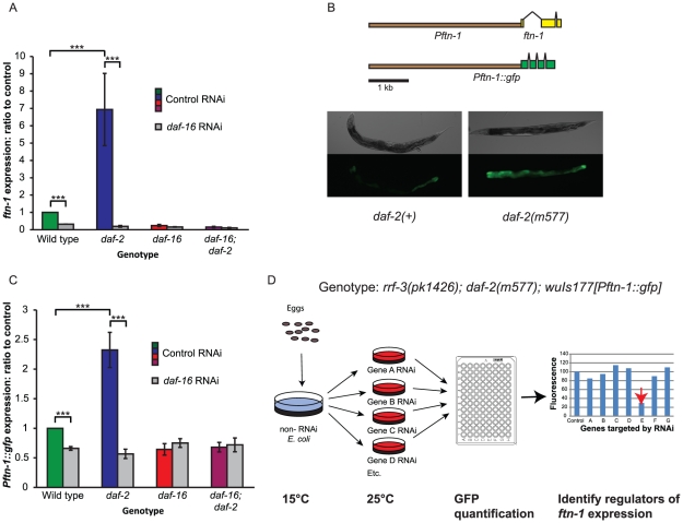
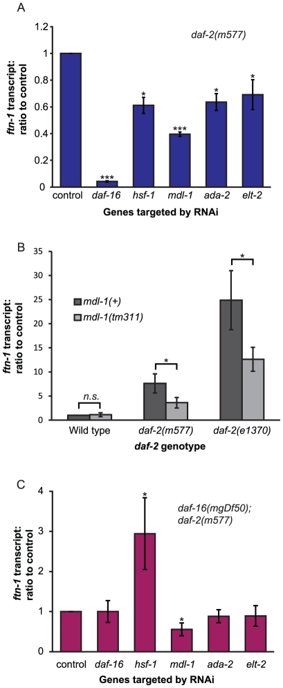
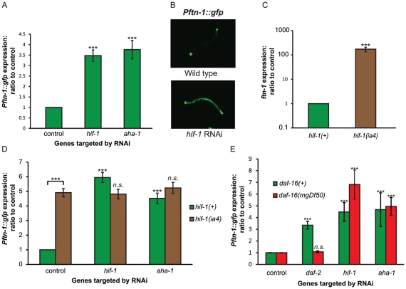
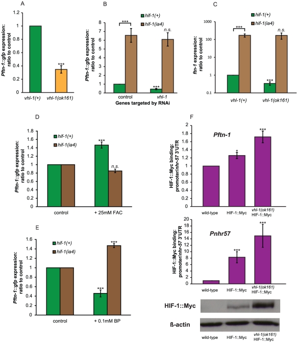
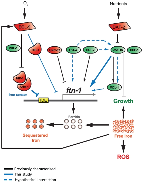
Iron plays an essential role in many biological processes, but also catalyzes the formation of reactive oxygen species (ROS), which can cause molecular damage. Iron homeostasis is therefore a critical determinant of fitness. In Caenorhabditis elegans, insulin/IGF-1 signaling (IIS) promotes growth and reproduction but limits stress resistance and lifespan through inactivation of the DAF-16/FoxO transcription factor (TF). We report that long-lived daf-2 insulin/IGF-1 receptor mutants show a daf-16-dependent increase in expression of ftn-1, which encodes the iron storage protein H-ferritin. To better understand the regulation of iron homeostasis, we performed a TF-limited genetic screen for factors influencing ftn-1 gene expression. The screen identified the heat-shock TF hsf-1, the MAD bHLH TF mdl-1, and the putative histone acetyl transferase ada-2 as activators of ftn-1 expression. It also revealed that the HIFα homolog hif-1 and its binding partner aha-1 (HIFβ) are potent repressors of ftn-1 expression. ftn-1 expression is induced by exposure to iron, and we found that hif-1 was required for this induction. In addition, we found that the prolyl hydroxylase EGL-9, which represses HIF-1 via the von Hippel-Lindau tumor suppressor VHL-1, can also act antagonistically to VHL-1 in regulating ftn-1. This suggests a novel mechanism for HIF target gene regulation by these evolutionarily conserved and clinically important hydroxylases. Our findings imply that the IIS and HIF pathways act together to regulate iron homeostasis in C. elegans. We suggest that IIS/DAF-16 regulation of ftn-1 modulates a trade-off between growth and stress resistance, as elevated iron availability supports growth but also increases ROS production.


Conflict of interest statement

The authors have declared that no competing interests exist.

Figures

Figure 1
Figure 1. Regulation of the ferritin gene ftn-1 by insulin/IGF-1 signaling.
(A) Effect of loss of daf-2 and daf-16 function on ftn-1 mRNA levels. Animals were grown at 15°C until the L4 stage of development, then kept at 25°C for two days prior to RNA extraction. (B) Construction of Pftn-1::gfp transgenic C. elegans. Approximately 3.8 kb of upstream sequence was fused to the GFP coding sequence. Epifluorescence images of nematodes bearing wuIs177 [Pftn-1::gfp] in daf-2(+) or daf-2(m577) backgrounds. Animals were grown at 15°C until the L4 stage and then kept at 25°C for two days before microscopy. The same exposure time was used for both images. (C) Effect of loss of daf-2 and daf-16 function on Pftn-1::gfp expression (c.f. Figure 1A). Animals were grown at 15°C until the L4 stage of development and then transferred to RNAi plates. They were then kept at 25°C for two days before GFP fluorescence measurements. (D) Diagrammatic depiction of RNAi screening protocol. Eggs were isolated by alkaline hypochlorite treatment and synchronized populations were left to develop at 15°C until the L4 stage of development. L4 animals were transferred to RNAi plates and left at 25°C for two days. Quantification of GFP expression was carried out by picking 40 animals into microtitre plates and measuring fluorescence using a platereader with a GFP filter set. Statistical significance was calculated by ANOVA in all cases. ***: p<0.001.
Figure 2
Figure 2. Identification of genes activating ftn-1 expression.
(A) Effect of RNAi of several transcription factors on ftn-1 transcript levels in daf-2(m577) mutants. (B) Effect of loss of mdl-1 on ftn-1 expression in both daf-2(m577) and daf-2(e1370) mutants. Protocol as described for (A) above. (C) Effect of RNAi on ftn-1 transcript levels in daf-16(mgDf50); daf-2(m577) mutants. For all trials, samples were collected on day 2 of adulthood. Statistical significance calculated by ANOVA. n.s:. non-significant, *: p<0.05, ***: p<0.001.
Figure 3
Figure 3. HIF signaling regulates ftn-1 expression.
(A) Effect of RNAi of hif-1 and aha-1 on the expression of Pftn-1::gfp. This result was obtained from animals carrying wuIs177 [Pftn-1::gfp]. Animals were grown at 20°C, transferred to RNAi at the L4 stage and GFP fluorescence quantified two days later. (B) Epifluorescence image of Pftn-1::gfp under control conditions (L4440) and hif-1 RNAi. Animals were grown at 20°C and photographed on day 2 of adulthood. (C) Effect of hif-1(ia4) on ftn-1 transcript levels. Animals were grown at 20°C and samples were collected on day 1 of adulthood. (D) Effect of hif-1 and aha-1 RNAi on ftn-1 expression in wild-type and hif-1(ia4) animals. (E) Effect of hif-1, aha-1 and daf-16 RNAi on Pftn-1::gfp expression in wild type and daf-16 mutants. These measurements were carried out on L4 animals kept at 25°C. In order to quantify GFP expression in L4 animals, 60 rather than 40 animals were transferred to each well of the microtitre plates. Statistical significance calculated by ANOVA. n.s:. non-significant, ***: p<0.001.
Figure 4
Figure 4. Evidence that the HIF pathway acts as an iron sensor.
(A) Effect vhl-1(ok161) on Pftn-1::gfp expression. Experiment was carried out at 20°C. (B) Effect of RNAi of vhl-1 on Pftn-1::gfp expression in wild-type and hif-1(ia4) animals. Nematodes were maintained on RNAi plates for two generations at 25°C and GFP fluorescence quantified at the L4 stage. (C) ftn-1 transcript levels in wild type, vhl-1(ok161), hif-1(ia4) and hif-1(ia4); vhl-1(ok161) double mutants. Cultures were grown at 20°C and samples were collected at day 1 of adulthood. (D) Effect of addition of iron (25 mM FAC) on expression of Pftn-1::gfp in wild type and hif-1(ia4) mutants. (E) Effect of addition of 0.1 mM bipyridyl (BP) on expression of Pftn-1::gfp in wild type and hif-1(ia4) mutants. (F) Chromatin immunoprecipitation (ChIP) was carried out using N2 (wild type), ZG429 (hif-1::Myc) and GA654 (hif-1::Myc vhl-1(ok161). Binding was assessed by qRT-PCR of ChIP samples using primers against the promoters of ftn-1 and the known HIF-1 target gene nhr-57. Values obtained were normalized to using qRT-PCR with primers against the 3′UTR of nhr-57, to which HIF-1::Myc is not thought to bind. HIF-1::Myc protein levels were quantified by Western blot using the same antibody aliquot as used for the ChIP experiment. Statistical significance calculated by ANOVA. *: p<0.05, ***: p<0.001.
Figure 5
Figure 5. HIF-1 represses expression via the iron-dependent element (IDE).
(A) Expression of Pftn-1::gfp in wild-type and vhl-1(ok161) animals with or without addition of iron (25 mM FAC). GFP fluorescence was quantified using a plate reader after 18 h of iron treatment. At least ten biological replicates were quantified. Asterisks denote statistically significant difference when compared to non-iron treated controls. (B) Expression of Pftn-1::gfp in wild-type and vhl-1(ok161) animals with or without iron chelation (0.1 mM bipyridyl, BP). GFP fluorescence was quantified using a plate reader after 18 h of iron chelation. BP-treated animals were compared to ethanol control treated ones. (C) Effect of hif-1 RNAi on the Pftn-1::gfp transgene with or without the IDE regulatory element. Fluorescence was measured in L4 animals grown at 25°C through pixel density quantification of epifluorescence microscopy images. hif-1 RNAi was administered for one generation. Strains used: XA6900 and XA6902. (D) ide::gfp expression in hif-1(+) and hif-1(ia4) animals with or without the addition of iron. Fluorescence measured as in (C). FAC treatment was administered from egg to L4 stage of development. (E) ide::gfp expression in hif-1(+) and hif-1(ia4) animals with or without the addition of iron chelator. Due to toxic effects of BP treatment during development, BP treatment was administered for 18 h during adulthood. Quantification was carried out on the second day of adulthood using pixel density quantification. n.s:. non-significant, *: p<0.05, ***: p<0.001.
Figure 6
Figure 6. Regulation of ftn-1 expression by EGL-9.
(A) Effect of egl-9(sa307) deletion mutaion on Pftn-1::gfp expression. Epifluorescence microscopy and plate reader quantification of GFP fluorescence was carried out on day 2 of adulthood. (B) Effect of egl-9 deletion on ftn-1 transcript levels in hif-1(+) and hif-1(ia4) animals. Samples were collected at day 1 of adulthood. Statistical significance calculated by ANOVA. n.s:. non-significant, ***: p<0.001.
Figure 7
Figure 7. ftn-1 expression is regulated by both the insulin/IGF-1 and hypoxia signaling pathways.
This figure provides a diagrammatic representation of the gene regulatory networking controlling ftn-1 expression. It includes previously established regulatory elements (black lines), newly established regulatory elements (blue lines) and new, hypothetical regulatory elements (dashed blue lines). We identified a positive regulatory role for the genes mdl-1, hsf-1, ada-2 and daf-16. In the case of mdl-1, previous work suggests that this transcription factor acts downstream of DAF-16, but it is unclear whether this is true for ftn-1 regulation (hence second line, dotted blue, from DAF-16 to MDL-1), or whether MDL-1 acts independently of DAF-16 in this case. Loss of ada-2 or elt-2 reduces ftn-1 expression but we were unable to detect an effect of ada-2 or elt-2 RNAi in the absence of DAF-16. While this may be caused by a lack of sensitivity in our assay, it could also indicate that these factors may act together with DAF-16 or upstream of DAF-16 to regulate ftn-1 expression. We found that hif-1 and aha-1 repress ftn-1 expression and that hif-1 is required for iron-dependent regulation of ftn-1, implying that HIF acts as an iron sensor in C. elegans. However, HIF-1 activity on ftn-1 expression can be regulated through both vhl-1-dependent and independent pathways and our data shows that these pathways act antagonistically on ftn-1 expression. The VHL-1-independent inhibition of ftn-1 expression by EGL-9 could either involve activation of transcriptional repression by HIF-1 or (more parsimoniously) inhibition of transcriptional activation by HIF-1. The latter interpretation would suggest the presence of a co-regulator that turns HIF-1 into a transcriptional activator of ftn-1.

References

    1. Riddle DL, Albert PS. 1997. Genetic and Environmental Regulation of Dauer Larva Development.
    1. Hu PJ. Dauer. WormBook; 2007. pp. 1–19. - PMC - PubMed
    1. Cassada RC, Russell RL. The dauerlarva, a post-embryonic developmental variant of the nematode Caenorhabditis elegans. Developmental Biology. 1975;46:326–342. - PubMed
    1. Lithgow GJ, Walker GA. Stress resistance as a determinate of C. elegans lifespan. Mechanisms of Ageing and Development. 2002;123:765–771. - PubMed
    1. Henis-Korenblit S, Zhang P, Hansen M, McCormick M, Lee SJ, et al. Insulin/IGF-1 signaling mutants reprogram ER stress response regulators to promote longevity. Proc Natl Acad Sci U S A. 2010;107:9730–9735. - PMC - PubMed

Publication types

MeSH terms