. 2012;7(3):e32542.
doi: 10.1371/journal.pone.0032542. Epub 2012 Mar 1.

Chemotherapeutics and radiation stimulate MHC class I expression through elevated interferon-beta signaling in breast cancer cells

Affiliations

Chemotherapeutics and radiation stimulate MHC class I expression through elevated interferon-beta signaling in breast cancer cells

Shan Wan et al. PLoS One. 2012.

Abstract

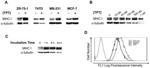
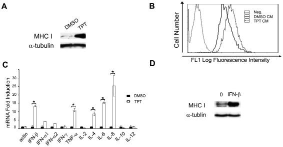
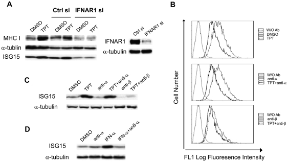
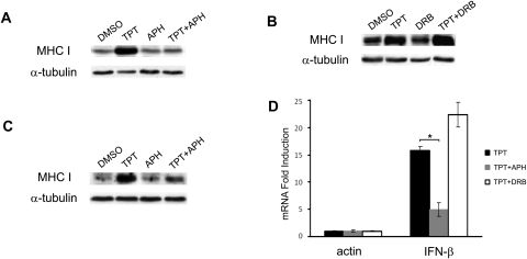
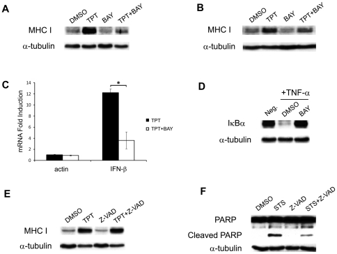
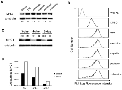
Low doses of anticancer drugs have been shown to enhance antitumor immune response and increase the efficacy of immunotherapy. The molecular basis for such effects remains elusive, although selective depletion of T regulatory cells has been demonstrated. In the current studies, we demonstrate that topotecan (TPT), a topoisomerase I-targeting drug with a well-defined mechanism of action, stimulates major histocompatibility complex class I (MHC I) expression in breast cancer cells through elevated expression/secretion of interferon-β (IFN-β) and activation of type I IFN signaling. First, we show that TPT treatment elevates the expression of both total and cell-surface MHC I in breast cancer cells. Second, conditioned media from TPT-treated breast cancer ZR-75-1 cells induce elevated expression of cell-surface MHC I in drug-naïve recipient cells, suggesting the involvement of cytokines and/or other secreted molecules. Consistently, TPT-treated cells exhibit elevated expression of multiple cytokines such as IFN-β, TNF-α, IL-6 and IL-8. Third, either knocking down the type I interferon receptor subunit 1 (IFNAR1) or addition of neutralizing antibody against IFN-β results in reduced MHC I expression in TPT-treated cells. Together, these results suggest that TPT induces increased IFN-β autocrine/paracrine signaling through type I IFN receptor, resulting in the elevated MHC I expression in tumor cells. Studies have also demonstrated that other chemotherapeutic agents (e.g. etoposide, cisplatin, paclitaxel and vinblastine) similarly induce increased IFN-β secretion and elevated MHC I expression. In addition, conditioned media from γ-irradiated donor cells are shown to induce IFN-β-dependent MHC I expression in unirradiated recipient cells. In the aggregate, our results suggest that many cancer therapeutics induce elevated tumor antigen presentation through MHC I, which could represent a common mechanism for enhanced antitumor immune response through T cell cytotoxicity during metronomic chemotherapy, as well as increased efficacy of combined chemo- (or radio-)/immuno-therapy.


Conflict of interest statement

Competing Interests: Sidney Pestka and Ronald Jubin are employees of PBL InterferonSource in the R & D Department. There are no patents, products in development, or marketed products to declare. This does not alter the authors' adherence to all the PLoS ONE policies on sharing data and materials, as detailed online in the guide for authors.

Figures

Figure 1
Figure 1. TPT induces elevated expression of both total and cell-surface MHC I in breast cancer cells.
(A) TPT induces elevated expression of total cellular MHC I in breast cancer cells. Breast cancer cell lines ZR-75-1, T47D, MDA-MB-231 (MB-231) and MCF-7 were treated with TPT (40, 200, 20, and 40 nM, respectively) for 4 days. Expression of total cellular MHC I was measured by immunoblotting using a polyclonal antibody against MHC I. (B) The concentration effect of TPT on total cellular expression of MHC I. ZR-75-1 cells were treated with increasing concentrations of topotecan (10 nM to 1 µM) for 4 days, followed by immunoblotting for total cellular MHC I expression. (C) Acute (1 hr) TPT exposure stimulates total cellular expression of MHC I. ZR-75-1 cells were treated with TPT (40 nM) for increasing durations of time (0, 1, 6 and 24 hrs), followed by incubation in drug-free medium for a total combined incubation of 4 days. Total cellular expression of MHC I was measured by immunoblotting. (D) TPT induces elevated cell-surface expression of MHC I. ZR-75-1 cells were treated with 40 nM of TPT for 1 hr, followed by incubation in drug-free medium for 4 days. Cell-surface MHC I was measured by FACS analysis using the pan-MHC I monoclonal antibody, W6/32.
Figure 2
Figure 2. Increased secretion of IFN-β is responsible for TPT-induced MHC I expression.
(A) Conditioned media from TPT-treated cells stimulate total cellular expression of MHC I in drug-naïve cells. ZR-75-1 cells were treated with 40 nM TPT for 1 hr, followed by incubation in drug-free medium for 4 days. Drug-naïve ZR-75-1 cells were then replenished with the conditioned media for another 2-day incubation. The total expression of cellular MHC I in recipient cells was measured by immunoblotting. (B) Conditioned media from TPT-treated cells stimulate cell-surface expression of MHC I in drug-naïve recipient cells. Recipient ZR-75-1 cells were treated with the conditioned media as described above, and the cell-surface MHC I expression was measured by FACS analysis. (C) TPT treatment increases mRNA levels of various cytokines in ZR-75-1 cells. Cells were treated with TPT (40 nM) for 1 hr, followed by incubation in drug-free medium for 3 days. Total RNAs were harvested for analysis by real-time RT-PCR. The experiments have been repeated twice. *: p-value<0.05. The error bar indicates standard deviation. (D) Purified IFN-β induces total cellular expression of MHC I. ZR-75-1 cells were treated with recombinant human IFN-β (500 U/ml) for 2 days. The total expression of cellular MHC I in recipient cells was measured by immunoblotting.
Figure 3
Figure 3. IFN-β signaling through type I IFN receptor is responsible for the TPT-induced MHC I expression.
(A) IFNAR1 knockdown abolishes TPT-induced total cellular MHC I expression. On the left panel, ZR-75-1 cells were mock-transfected, or transfected with control siRNA or IFNAR1-specific siRNA. 48 hrs post-transfection, cells were treated with TPT (or 0.1% DMSO as control) for 1 hr, followed by incubation in drug-free medium for 4 days. Lysates were analyzed by immunoblotting. On the right panel, ZR-75-1 cells were transfected with control or IFNAR1-specific siRNA. 72 hrs post-transfection, cells were harvested and immunoblotted with an anti-IFNAR1 antibody. (B) Neutralizing antibody against IFN-β, but not IFN-α, blocks the TPT-induced cell-surface MHC I expression. ZR-75-1 cells were treated with TPT (40 nM or 0.1% DMSO as control) for 1 hr, followed by incubation in drug-free medium with or without neutralizing antibody against IFN-α (anti-α, 1.44×10³ NU/ml) or IFN-β (anti-β, 296 NU/ml). Cell-surface MHC I expression was measured by FACS analysis. (C) Neutralizing antibody against IFN-β, but not IFN-α, blocks the TPT-induced ISG15. Antibody and TPT treatments of ZR-75-1 cells were performed exactly the same as described in (B). Lysates were immunoblotted for ISG15 expression. (D) Neutralizing antibody against IFN-α inhibits IFN-α-induced ISG15 expression. ZR-75-1 cells were co-incubated with IFN-α (100 U/ml) in the presence or absence of anti-IFN-α antibody (1.44×10³ NU/ml) for 2 days. Cells were then harvested and immunoblotted with an anti-ISG15 antibody.
Figure 4
Figure 4. TPT-induced elevation of MHC I and IFN-β is DNA replication-, but not transcription-, dependent.
(A) TPT-induced MHC I expression require active DNA synthesis. ZR-75-1 cells were pre-treated with APH (10 µM) for 30 min, followed by 0.1% DMSO or TPT (40 nM) treatment for 1 hr, and subsequent incubation in drug-free medium for 4 days. Expression of MHC I was then measured by immunoblotting. (B) TPT-induced MHC I expression is independent of transcription. ZR-75-1 cells were pre-treated with the transcription inhibitor DRB (150 µM) for 30 min, and then treated with TPT as described in (A). Expression of MHC I was measured by immunoblotting. (C) Conditioned medium-induced MHC I expression in recipient cells requires active DNA synthesis in TPT-treated donor cells. Recipient ZR-75-1 cells were incubated with conditioned media from TPT-treated donor cells as described in (A). The total expression of cellular MHC I in recipient cells was then measured by immunoblotting. (D) TPT-induced IFN-β mRNA expression requires active DNA synthesis. ZR-75-1 cells were pretreated with APH (10 µM) or DRB (150 µM) for 30 min prior to co-incubation with TPT (40 nM, 1 hr), followed by drug-free incubation for 3 days. Total RNAs were isolated for real-time RT-PCR analysis. The experiments have been repeated three times. *: p-value<0.05. The error bar indicates standard deviation.
Figure 5
Figure 5. TPT-induced MHC I expression requires NF-κB activation, but not apoptotic caspases.
(A) The NF-κB inhibitor BAY 11-7085 (BAY) blocks TPT-induced MHC I expression. ZR-75-1 cells were pretreated with BAY (10 µM) for 30 min, followed by co-incubation with 0.1% DMSO or TPT (40 nM) for 1 hr, and subsequent drug-free incubation for 4 days. Total cellular MHC I was then analyzed by immunoblotting. (B) The conditioned medium-induced MHC I in recipient cells requires NF-κB activation in TPT-treated donor cells. Drug- naïve recipient ZR-75-1 cells were incubated with the conditioned medium from TPT-treated donor cells as described in (A). Expression of total cellular MHC I in recipient cells was then analyzed by immunoblotting. (C) TPT-induced IFN-β mRNA expression requires NF-κB activation. ZR-75-1 cells were pretreated with BAY (10 µM) for 30 min prior to co-incubation with TPT (40 nM, 1 hr), followed by drug-free incubation for 3 days. Total RNAs were isolated for real-time RT-PCR analysis. The experiments have been repeated three times. *: p-value <0.05. The error bar indicates standard deviation. (D) BAY specifically blocks IκBα degradation induced by TNF-α treatment. ZR-75-1 cells were treated with TNF-α (10 ng/ml) for 10 min, in the presence or absence of BAY (10 µM). Expression of IκBα was measured by immunoblotting. (E) TPT-induced MHC I expression is independent of caspase activation. ZR-75-1 cells were pretreated with the pan-caspase inhibitor Z-VAD-FMK (Z-VAD, 10 µM) for 1 hr prior to co-incubation with TPT (40 nM, 1 hr), followed by continued incubation in drug-free medium for 4 days. Cell lysates were then immunoblotted with the anti-MHC I antibody. (F) Staurosporine-induced PAPR-1 cleavage requires caspase activation. ZR-75-1 cells were pretreated with Z-VAD-FMK (10 µM) for 1 hr followed by co-incubation with staurosporine (STS, 0.5 µM) for 6 hrs. PARP-1 cleavage was then measured by immunoblotting.
Figure 6
Figure 6. Cancer chemotherapeutics and ionizing radiation induce MHC I expression in breast cancer cells.
(A) Cancer chemotherapeutics induce elevated expression of total cellular MHC I in breast cancer ZR-75-1 cells. Cells were treated with topotecan (40 nM), etoposide (1 µM), cisplatin (6 µM), paclitaxel (3 µM), and vinblastine (6 nM) for 24 hrs, followed by incubation in drug-free medium for 3 days. Expression of total cellular MHC I was measured by immunoblotting. (B) Cancer chemotherapeutics induce cell-surface expression of MHC I in ZR-75-1 cells. ZR-75-1 cells were treated with different anticancer drugs as described in (A). Expression of cell-surface MHC I was then determined by FACS analysis. (C) Conditioned media from γ-irradiated cells induce elevated total cellular expression of MHC I in radiation-naïve recipient cells. Donor (ZR-75-1) cells were irradiated with γ-ray (2 Gy) for 3, 4, and 5 times. The conditioned media were then transferred to radiation-naïve recipient (ZR-75-1) cells. After 48 hrs, cell lysates were harvested for immunoblotting analysis. (D) Induction of MHC I expression in drug naïve recipient cells depends on IFN-β in the conditioned medium from irradiated donor cells. Radiation-naïve recipient (ZR-75-1) cells were incubated with the conditioned medium from irradiated (2 Gy×4) donor (ZR-75-1) cells in the presence of the neutralizing antibody against IFN-α (anti-α) or IFN-β (anti-β) for two days. Cell-surface MHC I in the recipient (ZR-75-1) cells was then measured by FACS analysis.

References

    1. Emens LA, Jaffee EM. Leveraging the Activity of Tumor Vaccines with Cytotoxic Chemotherapy. Cancer Research. 2005;65:8059–8064. - PubMed
    1. Pasquier E, Kavallaris M, Andre N. Metronomic chemotherapy: new rationale for new directions. Nat Rev Clin Oncol. 2010;7:455–465. - PubMed
    1. Emens LA. Chemoimmunotherapy. Cancer J. 2010;16:9. - PMC - PubMed
    1. Machiels J-PH, Reilly RT, Emens LA, Ercolini AM, Lei RY, et al. Cyclophosphamide, Doxorubicin, and Paclitaxel Enhance the Antitumor Immune Response of Granulocyte/Macrophage-Colony Stimulating Factor-secreting Whole-Cell Vaccines in HER-2/neu Tolerized Mice. Cancer Research. 2001;61:3689–3697. - PubMed
    1. Salem ML, Kadima AN, El-Naggar SA, Rubinstein MP, Chen Y, et al. Defining the ability of cyclophosphamide preconditioning to enhance the antigen-specific CD8+ T-cell response to peptide vaccination: creation of a beneficial host microenvironment involving type I IFNs and myeloid cells. J Immunother. 2007;30:14. - PubMed

Publication types

MeSH terms