

Quantum dot enabled detection of Escherichia coli using a cell-phone
- PMID: 22396952
- PMCID: PMC3683133
- DOI: 10.1039/c2an35071h
Quantum dot enabled detection of Escherichia coli using a cell-phone
Abstract
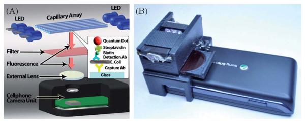
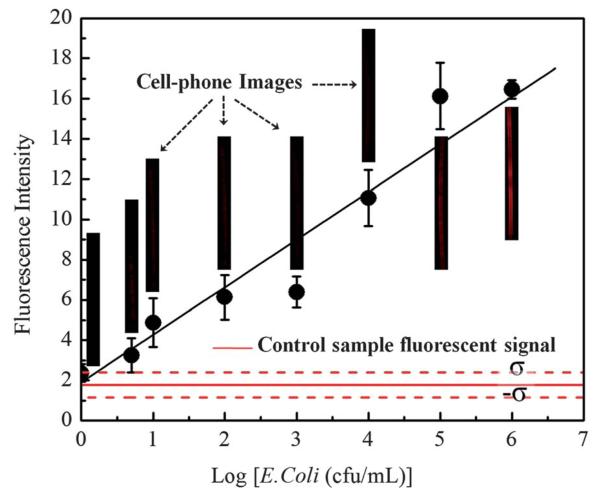
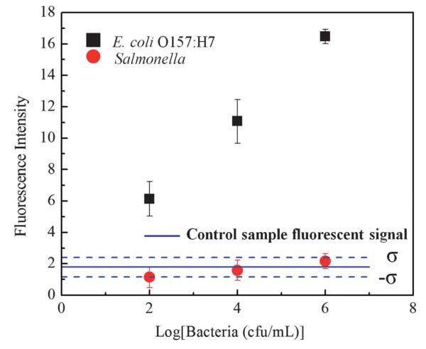
We report a cell-phone based Escherichia coli (E. coli) detection platform for screening of liquid samples. In this compact and cost-effective design attached to a cell-phone, we utilize anti-E. coli O157:H7 antibody functionalized glass capillaries as solid substrates to perform a quantum dot based sandwich assay for specific detection of E. coli O157:H7 in liquid samples. Using battery-powered inexpensive light-emitting-diodes (LEDs) we excite/pump these labelled E. coli particles captured on the capillary surface, where the emission from the quantum dots is then imaged using the cell-phone camera unit through an additional lens that is inserted between the capillary and the cell-phone. By quantifying the fluorescent light emission from each capillary tube, the concentration of E. coli in the sample is determined. We experimentally confirmed the detection limit of this cell-phone based fluorescent imaging and sensing platform as ∼5 to 10 cfu mL(-1) in buffer solution. We also tested the specificity of this E. coli detection platform by spiking samples with different species (e.g., Salmonella) to confirm that non-specific binding/detection is negligible. We further demonstrated the proof-of-concept of our approach in a complex food matrix, e.g., fat-free milk, where a similar detection limit of ∼5 to 10 cfu mL(-1) was achieved despite challenges associated with the density of proteins that exist in milk. Our results reveal the promising potential of this cell-phone enabled field-portable and cost-effective E. coli detection platform for e.g., screening of water and food samples even in resource limited environments. The presented platform can also be applicable to other pathogens of interest through the use of different antibodies.
Figures

References
-
- Dziuban EJ, Liang JL, Graun GF, Hill V, Painter J, Moore MR, Calderon RL, Roy SL, Beach MJ. Surveillance Summaries. 2006;55:1–24. - PubMed
-
- Shannon MA, Bohn PW, Elimelech M, Georgiadis JG, Marinas BJ, Mayes AM. Nature. 2008;452:301–310. - PubMed
-
- Fenwick A. Science. 2006;313:1077. - PubMed
-
-
Waterborne disease could cost over $ 500 million annually in U.S., http://www.cdc.gov/media/pressrel/2010/r100714.htm.
-
-
- Marris E. Nature. 2008;452:288. - PubMed
Publication types
MeSH terms
Substances
Grants and funding
LinkOut - more resources
Full Text Sources
Other Literature Sources