. 2012 Mar 7:10:24.
doi: 10.1186/1741-7015-10-24.

Fasudil improves survival and promotes skeletal muscle development in a mouse model of spinal muscular atrophy

Affiliations

Fasudil improves survival and promotes skeletal muscle development in a mouse model of spinal muscular atrophy

Melissa Bowerman et al. BMC Med. .

Abstract

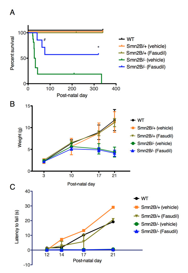
Background: Spinal muscular atrophy (SMA) is the leading genetic cause of infant death. It is caused by mutations/deletions of the survival motor neuron 1 (SMN1) gene and is typified by the loss of spinal cord motor neurons, muscular atrophy, and in severe cases, death. The SMN protein is ubiquitously expressed and various cellular- and tissue-specific functions have been investigated to explain the specific motor neuron loss in SMA. We have previously shown that the RhoA/Rho kinase (ROCK) pathway is misregulated in cellular and animal SMA models, and that inhibition of ROCK with the chemical Y-27632 significantly increased the lifespan of a mouse model of SMA. In the present study, we evaluated the therapeutic potential of the clinically approved ROCK inhibitor fasudil.

Methods: Fasudil was administered by oral gavage from post-natal day 3 to 21 at a concentration of 30 mg/kg twice daily. The effects of fasudil on lifespan and SMA pathological hallmarks of the SMA mice were assessed and compared to vehicle-treated mice. For the Kaplan-Meier survival analysis, the log-rank test was used and survival curves were considered significantly different at P < 0.05. For the remaining analyses, the Student's two-tail t test for paired variables and one-way analysis of variance (ANOVA) were used to test for differences between samples and data were considered significantly different at P < 0.05.

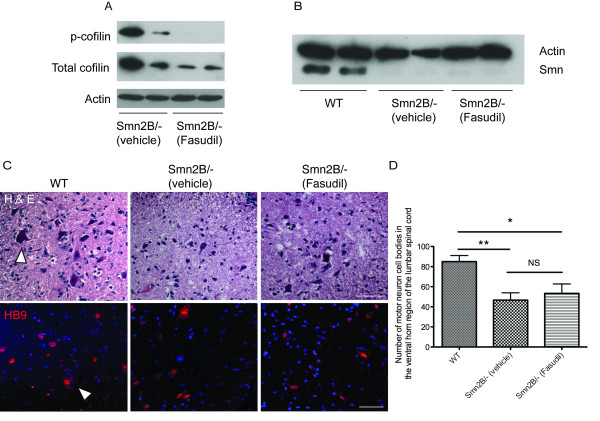
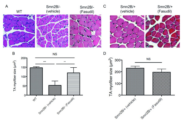
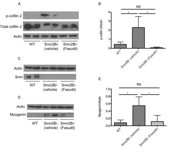
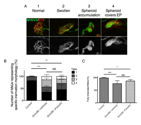
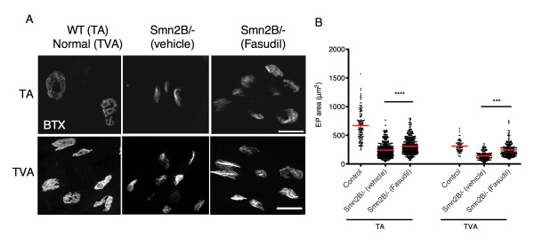
Results: Fasudil significantly improves survival of SMA mice. This dramatic phenotypic improvement is not mediated by an up-regulation of Smn protein or via preservation of motor neurons. However, fasudil administration results in a significant increase in muscle fiber and postsynaptic endplate size, and restores normal expression of markers of skeletal muscle development, suggesting that the beneficial effects of fasudil could be muscle-specific.

Conclusions: Our work underscores the importance of muscle as a therapeutic target in SMA and highlights the beneficial potential of ROCK inhibitors as a therapeutic strategy for SMA and for other degenerative diseases characterized by muscular atrophy and postsynaptic immaturity.


Figures

Figure 1
Figure 1
Fasudil increases lifespan of Smn2B/- mice, independent of weight gain and pen test performance. Fasudil (30 mg/kg twice daily) or vehicle (water) was administered by gavage from post-natal (P) day 3 to P21. The different groups analyzed were: untreated wild type (WT) (n = 10), vehicle-treated Smn2B/+ (n = 8), fasudil-treated Smn2B/+ (n = 9), vehicle-treated Smn2B/- (n = 16) and fasudil-treated Smn2B/- (n = 7). (A) Fasudil significantly increases lifespan of Smn2B/- mice when compared to vehicle-treated Smn2B/- mice (*P = 0.0251; # indicates death due to dystocia). Administration of fasudil does not have adverse effects on the lifespan of normal littermates. (B) Fasudil does not prevent the arrest in weight gain that occurs in vehicle-treated Smn2B/- mice onwards of P10.(C) Fasudil does not improve the performance of Smn2B/- mice on the pen test when compared to vehicle-treated Smn2B/- mice.
Figure 2
Figure 2
Fasudil activity in the spinal cord is Smn-independent and does not prevent motor neuron loss in the ventral horn region. (A and B) Spinal cords were obtained from post-natal (P) day 21 untreated wild type (WT), vehicle-treated Smn2B/- and fasudil-treated Smn2B/- mice. (A) Immunoblot analysis shows that spinal cords treated with the rho-kinase (ROCK) inhibitor fasudil have decreased levels of p-cofilin, a known substrate of ROCK. (B) Immunoblot analysis shows that fasudil does not increase Smn protein levels in the spinal cords of Smn2B/- mice. (C and D) Spinal cord sections were analyzed from P21 untreated wild type (WT) (n = 3), vehicle-treated Smn2B/- (n = 3) and fasudil-treated Smn2B/- (n = 3) mice. (C) Representative images of hematoxylin and esoin (H&E)- and HB9-stained spinal cord sections. Arrowhead depicts a typical large motor neuron. Scale bar = 50 μm. (D) Quantification of motor neurons within the ventral horn region of the spinal cord shows that fasudil does not prevent the motor neuron loss that occurs in vehicle-treated Smn2B/- mice (*P < 0.05; **P < 0.01; NS = not significant; data are mean +/- s.d.).
Figure 3
Figure 3
Fasudil increases tibialis anterior (TA) myofiber size. TA muscles were isolated from post-natal (P) day 21 untreated wild type (WT) (n = 3), vehicle-treated Smn2B/+ (n = 3), fasudil-treated Smn2B/+ (n = 3), vehicle-treated Smn2B/- (n = 6) and fasudil-treated Smn2B/- (n = 6) mice. (A) Representative images of cross-sections of WT, or vehicle- and fasudil-treated Smn2B/- TA muscles stained with hematoxylin and eosin. Scale bar = 50 μm. (B) Quantification shows that fasudil-treated Smn2B/- TA muscles display significantly larger myofibers than vehicle-treated Smn2B/- mice. (**P < 0.01; ***P < 0.001; NS = not significant; data are mean +/- s.d.). (C) Representative images of cross-sections of vehicle- and fasudil-treated Smn2B/+ TA muscles stained with hematoxylin and eosin. Scale bar = 50 μm. (D) Quantification shows that fasudil does not significantly increase the myofiber size of Smn2B/+ normal mice (NS = not significant; data are mean +/- s.d.).
Figure 4
Figure 4
Fasudil administration inhibits ROCK in skeletal muscle and restores normal myogenin expression levels. Tibialis anterior (TA) muscles were isolated from post-natal (P) day 21 untreated wild type (WT) (n = 4), vehicle-treated Smn2B/- (n = 4) and fasudil-treated Smn2B/- (n = 4) mice. (A) Immunoblot analysis of p-cofilin 2, a muscle-specific downstream substrate of ROCK, shows that its levels are increased in the TA muscles of Smn2B/- mice compared with wild type. Furthermore, this experiment shows that fasudil inhibits ROCK in the TA muscle of Smn2B/- mice, and reduces the p-cofilin 2 levels to wild type levels. (B) Quantification shows that wild type TA muscles have significantly less p-cofilin 2 than vehicle-treated Smn2B/- TA muscles, and that fasudil is active in skeletal muscle and restores p-cofilin 2 to normal levels. (*P < 0.05; NS = not significant; data are mean +/- s.d.). (C) Immunoblot analysis shows that fasudil does not increase Smn protein levels in the TA muscles of Smn2B/- mice. (D) Immunoblot analysis shows that fasudil results in a decrease in myogenin protein levels in fasudil-treated Smn2B/- TA muscles when compared to vehicle-treated Smn2B/- mice. (E) Wild type muscle has significantly less myogenin than vehicle-treated Smn2B/- TA muscles. Fasudil administration to Smn2B/- mice restores myogenin to normal levels (*P < 0.05; NS = not significant; data are mean +/- s.d.).
Figure 5
Figure 5
Fasudil does not improve pre-synaptic neuromuscular junction (NMJ) phenotype of Smn2B/- mice. Pre-synaptic morphology was analyzed in the transversus abdominis (TVA) muscle of post-natal (P) day 21 untreated control littermates (n = 3), vehicle-treated Smn2B/- (n = 6) and fasudil-treated Smn2B/- (n = 4) mice. (A) Representative images of NMJs depicting the pre-synaptic morphology categories: normal (type 1), swollen (type 2), spheroid accumulation (type 3) and spheroids covers the endplate (EP) (type 4). (Neurofilament (NF) and synaptic vesicle protein 2 (SV2): green; EP: red (BTX)). (B) Quantification of the pre-synaptic morphology shows that control littermates have significantly more 'normal' (type 1) NMJs than both vehicle- and fasudil-treated Smn2B/- mice (**P < 0.01; ***P < 0.001; NS = not significant; data are mean +/- s.d.). C) Quantification of the fully innervated NMJs shows that both vehicle- and fasudil-treated Smn2B/- muscles display significantly fewer fully innervated NMJs than control littermates, with no significant difference between vehicle or Ffsudil treated Smn2B/- mice (*P < 0.05; ***P < 0.001; NS = not significant; data are mean +/- s.d.). BTX, α-bungarotoxin.
Figure 6
Figure 6
Fasudil increases endplate (EP) area in the tibialis anterior (TA) and transversus abdominis (TVA) muscles. Muscles were isolated from post-natal (P) day 21 untreated control littermates (n = 3), vehicle-treated Smn2B/- (n = 4) and fasudil-treated Smn2B/- mice (n = 3). (A) Representative images of TA and TVA EPs stained with α-bungarotoxin (BTX). Scale bars = 25 μm (TA) and 30 μm (TVA). (B) Quantification of EP area shows that fasudil-treated Smn2B/- TA and TVA muscles display significantly larger EPs when compared to vehicle-treated Smn2B/- muscles. (***P < 0.001; ****P < 0.0001; data are mean +/- s.d.).
Figure 7
Figure 7
Aging fasudil-treated Smn2B/- mice display mature neuromuscular junctions (NMJs). Transversus abdominis (TVA) muscles were isolated from post-natal (P) day 21 (n = 6) and 6-month -old (n = 4) fasudil-treated Smn2B/- mice. (A) Representative images of TVA muscles from P21 and 6-month-old fasudil-treated Smn2B/- mice. (Neurofilament (NF) and synaptic vesicle protein 2 (SV2): green; EP: red (BTX)). Scale bar = 30 μm. B) Types of EP occupation were categorized and quantified as fully occupied (where the pre-synaptic terminal completely covers the EP), partial (where the pre-synaptic terminal partially covers the EP), or vacant (where no pre-synaptic terminal is present at an EP). Surviving 6-month-old fasudil-treated Smn2B/- mice display significantly more fully occupied EPs than P21 fasudil-treated Smn2B/- mice (**P = 0.0096; data are mean +/- s.d.). (C) Representative images of EP morphology categorization from mature to immature: pretzel, perforated, bright lines and uniform. EPs are visualized with BTX. (D) Quantification shows that 6-month-old fasudil-treated Smn2B/- mice display significantly more mature pretzel-shaped EPs than P21 fasudil-treated Smn2B/- mice (****P < 0.0001); data are mean +/- s.d.). BTX, α-bungarotoxin; EP, endplate.

Similar articles

Cited by

References

    1. Crawford TO, Pardo CA. The neurobiology of childhood spinal muscular atrophy. Neurobiol Dis. 1996;3:97–110. doi: 10.1006/nbdi.1996.0010. - DOI - PubMed
    1. Pearn J. Incidence, prevalence, and gene frequency studies of chronic childhood spinal muscular atrophy. J Med Genet. 1978;15:409–413. doi: 10.1136/jmg.15.6.409. - DOI - PMC - PubMed
    1. Lefebvre S, Burglen L, Reboullet S, Clermont O, Burlet P, Viollet L, Benichou B, Cruaud C, Millasseau P, Zeviani M. et al.Identification and characterization of a spinal muscular atrophy-determining gene. Cell. 1995;80:155–165. doi: 10.1016/0092-8674(95)90460-3. - DOI - PubMed
    1. Jablonka S, Schrank B, Kralewski M, Rossoll W, Sendtner M. Reduced survival motor neuron (Smn) gene dose in mice leads to motor neuron degeneration: an animal model for spinal muscular atrophy type III. Hum Mol Genet. 2000;9:341–346. doi: 10.1093/hmg/9.3.341. - DOI - PubMed
    1. Lorson CL, Hahnen E, Androphy EJ, Wirth B. A single nucleotide in the SMN gene regulates splicing and is responsible for spinal muscular atrophy. Proc Natl Acad Sci USA. 1999;96:6307–6311. doi: 10.1073/pnas.96.11.6307. - DOI - PMC - PubMed

Publication types

MeSH terms