Review
. 2012 Mar 7:12:11.
doi: 10.1186/1471-2377-12-11.

Meningeal and cortical grey matter pathology in multiple sclerosis

Affiliations
Review

Meningeal and cortical grey matter pathology in multiple sclerosis

Bogdan F Gh Popescu et al. BMC Neurol. .

Abstract

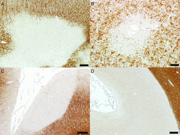
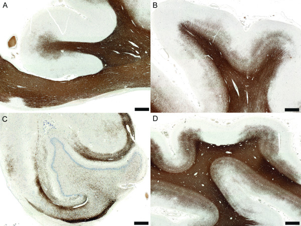
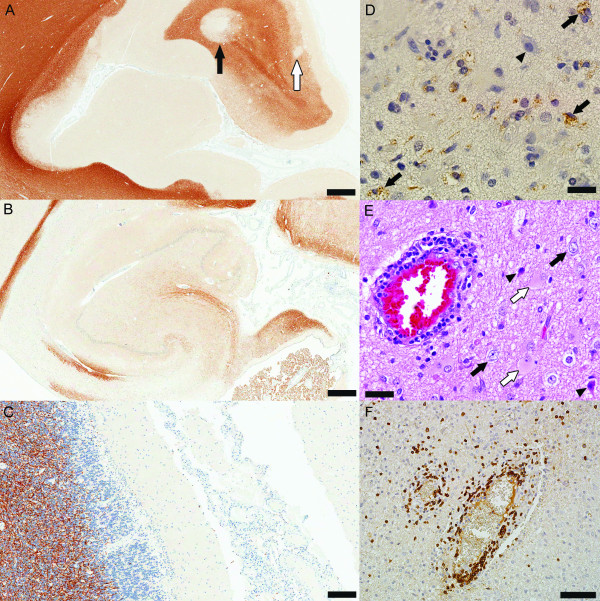
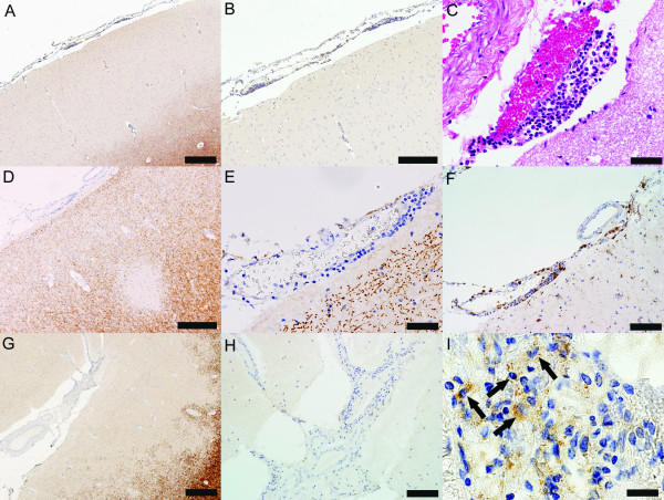
Although historically considered a disease primarily affecting the white matter of the central nervous system, recent pathological and imaging studies have established that cortical demyelination is common in multiple sclerosis and more extensive than previously appreciated. Subpial, intracortical and leukocortical lesions are the three cortical lesion types described in the cerebral and cerebellar cortices of patients with multiple sclerosis. Cortical demyelination may be the pathological substrate of progression, and an important pathologic correlate of irreversible disability, epilepsy and cognitive impairment. Cortical lesions of chronic progressive multiple sclerosis patients are characterized by a dominant effector cell population of microglia, by the absence of macrophagic and leukocytic inflammatory infiltrates, and may be driven in part by organized meningeal inflammatory infiltrates. Cortical demyelination is also present and common in early MS, is topographically associated with prominent meningeal inflammation and may even precede the appearance of classic white matter plaques in some MS patients. However, the pathology of early cortical lesions is different than that of chronic MS in the sense that early cortical lesions are highly inflammatory, suggesting that neurodegeneration in MS occurs on an inflammatory background and raising interesting questions regarding the role of cortical demyelination and meningeal inflammation in initiating and perpetuating the disease process in early MS.


Figures

Figure 1
Figure 1
Cortical demyelinated lesion types. (A) Leukocortical lesion (PLP, scale bar = 100 μm); (B) Intracortical lesion (PLP, scale bar = 500 μm); (C) Subpial lesion affecting the superficial cortical layers (PLP, scale bar = 250 μm); (D) Subpial lesion involving all cortical layers without affecting the white matter (PLP, scale bar = 500 μm).
Figure 2
Figure 2
Non-inflammatory cortical demyelination is prominent in chronic MS. (A) Cortical demyelination in the cingulate gyrus (PLP, scale bar = 5 mm); (B) Cortical demyelination in the frontal lobe (PLP, scale bar = 5 mm); (C) Cortical demyelination in the hippocampus (PLP, scale bar = 2.5 mm); (D) Although the occipital lobe is less frequently affected, cortical demyelination can be very extensive in the occipital lobe of some MS patients (PLP, scale bar = 5 mm).
Figure 3
Figure 3
Inflammatory cortical demyelination is present and common in early MS. (A-C) Tissue obtained at autopsy form patients with acute MS: (A) Prominent cortical demyelination in the neocortex with the presence of all cortical lesion types: extensive subpial demyelination, intracortical demyelination (white arrow) and leukocortical demyelination (black arrow) (PLP, scale bar = 2.5 mm); (B) Cortical demyelination in the hippocampus (PLP, scale bar = 2.5 mm); (C) Subpial cerebellar demyelinated lesion (PLP, scale bar = 500 μm). (D-E) Inflammatory cortical demyelination in the cortex from biopsied patients: (D) Demyelinated cortex (arrowhead indicates a neuron) infiltrated by myelin-laden macrophages consistent with active demyelination (arrows) (PLP; scale bar = 50 μm); (E) Perivascular inflammation, degenerating neurons (arrowheads) scattered among normal-looking neurons (black arrows) and reactive astrocytes (white arrows) are present in cortical lesions in early MS (HE, scale bar = 50 μm); (F) Lymphocytic perivascular and diffuse inflammatory infiltrates in the demyelinated cortex (CD3, scale bar = 250 μm).
Figure 4
Figure 4
Meningeal inflammation is topographically associated with cortical demyelination in early MS (autopsy tissue from patients with acute MS). (A) Subpial lesion overlaid by a perivascular meningeal inflammatory infiltrate (PLP, scale bar = 500 μm); (B) Higher magnification of (A) (PLP, scale bar = 250 μm); (C) Higher magnification of the perivascular meningeal infiltrate (HE, scale bar = 50 μm); (D) Intracortical lesion overlaid by perivascular meningeal inflammation (PLP, scale bar = 250 μm); (E) Higher magnification of the perivascular meningeal infiltrate (PLP, scale bar = 50 μm); (F) Macrophages are components of the perivascular meningeal infiltrate (KiM1P, scale bar = 100 μm); (G) Large subpial lesion underlying meningeal inflammation (PLP, scale bar = 500 μm); (H) Higher magnification of the meningeal inflammation (PLP, scale bar = 100 μm); (I) Myelin-laden macrophages are found in the meninges of patients with acute MS (PLP, scale bar = 20 μm).

References

    1. Anderson DW, Ellenberg JH, Leventhal CM, Reingold SC, Rodriguez M, Silberberg DH. Revised estimate of the prevalence of multiple sclerosis in the United States. Ann Neurol. 1992;31(3):333–336. doi: 10.1002/ana.410310317. - DOI - PubMed
    1. Charcot JM. Lectures on the Diseases of the Nervous System. London: The New Sydenham Society; 1887. Lecture VI. Disseminated Sclerosis. Pathological Anatomy; pp. 157–181.
    1. Sobel RA, Moore GRW. In: Greenfield's Neuropathology. Volume 2, 8th edition. Love S, Louis DN, Ellison DW, editor. Oxford University Press; 2008. Demyelinating diseases; pp. 1513–1608. - PubMed
    1. Peterson JW, Bo L, Mork S, Chang A, Trapp BD. Transected neurites, apoptotic neurons, and reduced inflammation in cortical multiple sclerosis lesions. Ann Neurol. 2001;50(3):389–400. doi: 10.1002/ana.1123. - DOI - PubMed
    1. Brownell B, Hughes JT. The distribution of plaques in the cerebrum in multiple sclerosis. J Neurol Neurosurg Psychiatry. 1962;25:315–320. doi: 10.1136/jnnp.25.4.315. - DOI - PMC - PubMed

Publication types