Review
. 2012 Mar 7:9:47.
doi: 10.1186/1742-2094-9-47.

Who fans the flames of Alzheimer's disease brains? Misfolded tau on the crossroad of neurodegenerative and inflammatory pathways

Affiliations
Review

Who fans the flames of Alzheimer's disease brains? Misfolded tau on the crossroad of neurodegenerative and inflammatory pathways

Norbert Zilka et al. J Neuroinflammation. .

Abstract

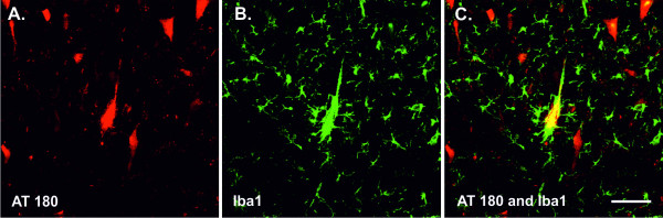
Neurodegeneration, induced by misfolded tau protein, and neuroinflammation, driven by glial cells, represent the salient features of Alzheimer's disease (AD) and related human tauopathies. While tau neurodegeneration significantly correlates with disease progression, brain inflammation seems to be an important factor in regulating the resistance or susceptibility to AD neurodegeneration. Previously, it has been shown that there is a reciprocal relationship between the local inflammatory response and neurofibrillary lesions. Numerous independent studies have reported that inflammatory responses may contribute to the development of tau pathology and thus accelerate the course of disease. It has been shown that various cytokines can significantly affect the functional and structural properties of intracellular tau. Notwithstanding, anti-inflammatory approaches have not unequivocally demonstrated that inhibition of the brain immune response can lead to reduction of neurofibrillary lesions. On the other hand, our recent data show that misfolded tau could represent a trigger for microglial activation, suggesting the dual role of misfolded tau in the Alzheimer's disease inflammatory cascade. On the basis of current knowledge, we can conclude that misfolded tau is located at the crossroad of the neurodegenerative and neuroinflammatory pathways. Thus disease-modified tau represents an important target for potential therapeutic strategies for patients with Alzheimer's disease.


Figures

Figure 1
Figure 1
Activated microglia are localized in brain areas affected by neurofibrillary tangles. Reactive microglia, stained by Iba1 antibody (red colour), are distributed throughout the brain regions affected by neurofibrillary lesions immunolabelled with AT8 (antibody recognizing phospho-tau, green colour) and DC11 (antibody recognizing Alzheimer's disease (AD)-specific misfolded tau, green colour). Activated microglia are surrounding tangle-bearing neurons in the hippocampus of an AD patient (A) and (B) and in the cortices of transgenic rats expressing truncated tau protein (C) and (D). Scale bars: 100 μm.
Figure 2
Figure 2
Colocalization of clustered microglia and neurofibrillary tangles in transgenic rat brain. Neurofibrillary tangles (NFTs) were detected with the monoclonal antibody AT180, recognizing tau protein phosphorylated at Thr231 and Ser235 (A), respectively. Activated microglia were stained with the polyclonal antibody Iba1, specific for ionized calcium-binding adaptor molecule 1 (B). Confocal study showed that some NFTs colocalized with clusters of activated microglia (C). Scale bars: 100 μm.
Figure 3
Figure 3
Functional plasticity of microglial cells in transgenic rat brain expressing human truncated tau protein. Intraneuronal expression of human truncated tau induces neurofibrillary degeneration. Injured neurons drive resting microglia to become reactive microglia that transform into brain phagocytic microglia: brain macrophages. At this stage, microglia lose contact inhibition and begin to fuse with each other, forming small clusters. Morphological activation of microglia is accompanied with upregulation of several immunological markers, including integrins CD11a, CD11b, CD11c and CD18; lymphocytic antigen CD4; leukocyte common antigen CD45; and lysosomal glycoprotein CD68 (colour dots). In the late stage of neurodegeneration, bloodborne leukocytes (mainly monocytes and partially dendritic cells) infiltrate the brain parenchyma and participate in the brain's immune response.
Figure 4
Figure 4
Tau neuroinflammatory cascade. In early stages of tau structural metamorphosis, some proinflammatory cytokines (IL-1, IL6 and TNF-α) and chemokines (fractalkine) can modify tau phosphorylation patterns and thus change the structure and function of tau protein. The major sources of proinflammatory cytokines are activated microglia and astrocytes. Glial cells can be activated by different inducers, including misfolded tau protein. At this stage, however, the phosphorylated tau protein would not necessarily aggregate into the filamentous structures (A). In later stages, when misfolded hyperphosphorylated tau proteins form mature NFTs, inflammation could either accelerate or modify tangle formation. Several components of the brain's immune system, including cytokines and proteins of the complement pathway, have been shown to be involved in the molecular dialogue between activated glial cells and tangle-bearing neurons (B).

Similar articles

Cited by

References

    1. Grundke-Iqbal I, Iqbal K, Quinlan M, Tung YC, Zaidi MS, Wisniewski HM. Microtubule-associated protein tau: a component of Alzheimer paired helical filaments. J Biol Chem. 1986;261:6084–6089. - PubMed
    1. Grundke-Iqbal I, Iqbal K, Tung YC, Quinlan M, Wisniewski HM, Binder LI. Abnormal phosphorylation of the microtubule-associated protein tau (tau) in Alzheimer cytoskeletal pathology. Proc Natl Acad Sci USA. 1986;83:4913–4917. doi: 10.1073/pnas.83.13.4913. - DOI - PMC - PubMed
    1. Glenner GG, Wong CW. Alzheimer's disease and Down's syndrome: sharing of a unique cerebrovascular amyloid fibril protein. Biochem Biophys Res Commun. 1984;122:1131–1135. doi: 10.1016/0006-291X(84)91209-9. - DOI - PubMed
    1. Wischik CM, Novak M, Thøgersen HC, Edwards PC, Runswick MJ, Jakes R, Walker JE, Milstein C, Roth M, Klug A. Isolation of a fragment of tau derived from the core of the paired helical filament of Alzheimer disease. Proc Natl Acad Sci USA. 1988;85:4506–4510. doi: 10.1073/pnas.85.12.4506. - DOI - PMC - PubMed
    1. Wischik CM, Novak M, Edwards PC, Klug A, Tichelaar W, Crowther RA. Structural characterization of the core of the paired helical filament of Alzheimer disease. Proc Natl Acad Sci USA. 1988;85:4884–4888. doi: 10.1073/pnas.85.13.4884. - DOI - PMC - PubMed

Publication types