

Review
doi: 10.1186/1477-7819-10-49.
Osteoblastoma of the rib with CT and MR imaging: a case report and literature review
Affiliations
- PMID: 22397553
- PMCID: PMC3320550
- DOI: 10.1186/1477-7819-10-49
Item in Clipboard
Review
Osteoblastoma of the rib with CT and MR imaging: a case report and literature review
World J Surg Oncol.
.
Abstract
Osteoblastoma is a rare bone tumor which is mostly found in the vertebral column and long bone. We describe a 59-year-old woman with osteoblastoma in the right fifth posterior segment of the rib, whose presenting symptoms were right back pain for two years and awakened at night. Chest computer tomography (CT) and thoracic spine magnetic resonance (MR) imaging findings included an expansile lesion of the right fifth rib and an ossified matrix. Surgical resection of the lesion confirmed a benign osteoblastoma. 12 months follow-up revealed disappearance of right back pain. Rib osteoblastoma in plain film has been described previously; however, to our knowledge this is the only case report emphasized in CT and MR imaging.
Figures
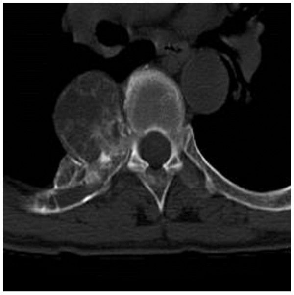
CT reveals a lytic lesion in right fifth posterior rib with ossified matrix.
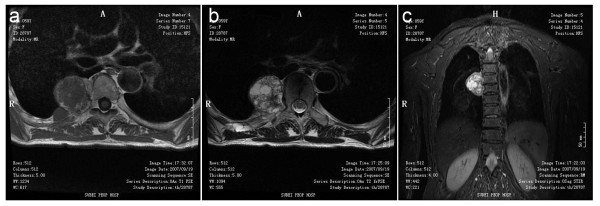
MR finding in right fifth posterior rib. MR imaging shows isointensity on a T1-weighted image (a), heterogeneous high signal pattern with a low signal rim on a T2-weighted image (b) and Coronal STIR imaging (c)
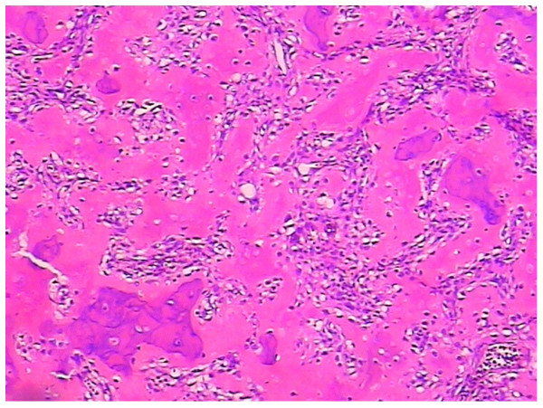
(Hematoxylin and eosin staining, ×100) shows irregular woven bone and osteoid surrounded by activated osteoblasts. Seven days after the operation the patient was discharged from hospital. At 12 months follow-up the preoperative symptoms disappeared completely and there were no post-operative complications.
References
-
- Cerase A, Priolo F. Skeletal benign bone-forming lesions. Eur J Radiol. 1998;27(Suppl 1):S91–S97. - PubMed
-
- Kroon HM, Schurmans J. Osteoblastoma: clinical and radiologic findings in 98 new cases. Radiology. 1990;175:783–790. - PubMed
-
- Jackson RP. Recurrent osteoblastoma: a review. Clin Orthop Relat Res. 1978;131:229–233. - PubMed
Publication types
MeSH terms
LinkOut - more resources
Full Text Sources
Medical
