Case Reports
. 2012 Mar 8;366(10):925-31.
doi: 10.1056/NEJMoa1112824.

Immunologic correlates of the abscopal effect in a patient with melanoma

Affiliations
Case Reports

Immunologic correlates of the abscopal effect in a patient with melanoma

Michael A Postow et al. N Engl J Med. .

Abstract

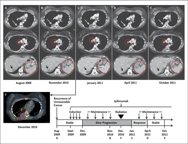
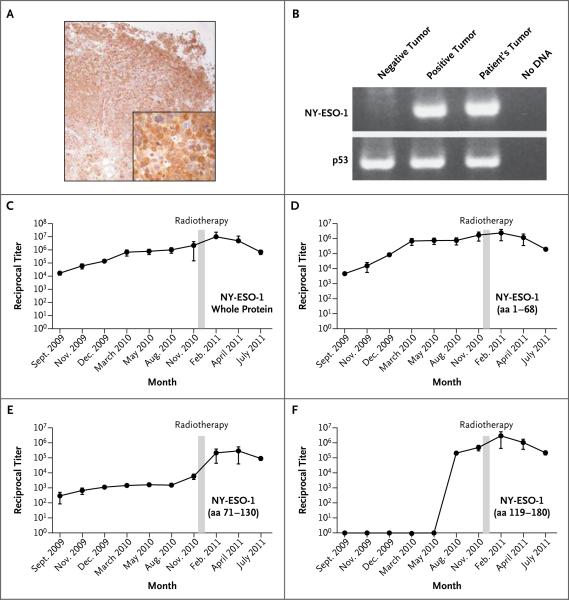
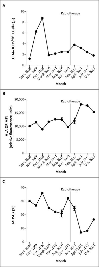
The abscopal effect is a phenomenon in which local radiotherapy is associated with the regression of metastatic cancer at a distance from the irradiated site. The abscopal effect may be mediated by activation of the immune system. Ipilimumab is a monoclonal antibody that inhibits an immunologic checkpoint on T cells, cytotoxic T-lymphocyte-associated antigen 4 (CTLA-4). We report a case of the abscopal effect in a patient with melanoma treated with ipilimumab and radiotherapy. Temporal associations were noted: tumor shrinkage with antibody responses to the cancer-testis antigen NY-ESO-1, changes in peripheral-blood immune cells, and increases in antibody responses to other antigens after radiotherapy. (Funded by the National Institutes of Health and others.).


Figures

Figure 1
Figure 1. Results of Diagnostic and Radiotherapy Simulation Imaging throughout the Disease Course
Axial CT images are shown, corresponding to the timeline showing therapy and disease status. White arrows indicate the paraspinal mass, red circles indicate the right hilar lymphadenopathy and spleen, and black arrows indicate an incidental hepatic hemangioma. Panel A (top) represents the status before treatment with ipilimumab. Panel B shows enlargement of the paraspinal mass (top), stable right hilar lymphadenopathy (middle), and new splenic lesions (bottom). Panel C shows images 1 month after radiotherapy, when the response to radiotherapy had not yet occurred, with apparent continued worsening disease at all three sites. Several months after radiotherapy, the targeted paraspinal mass showed a response (Panel D, top). Furthermore, disease response outside of the radiation field was seen with decreased right hilar lymphadenopathy (middle) and resolution of splenic lesions (bottom). The response was durable, as shown in Panel E. Panel F shows the CT simulation image for radiotherapy planning, with the target volume (indicated in purple) encompassing the right paraspinal metastatic mass. The isodose lines represent total doses of 2850 cGy (pink), 2000 cGy (orange), 1000 cGy (green), and 200 cGy (blue). Disease regression was confirmed by means of three-dimensional volumetric assessment (Table 2 in the Supplementary Appendix).
Figure 2
Figure 2. NY-ESO-1 Expression and Antibody Response to Ipilimumab and Radiotherapy
Immunohistochemical analysis of NY-ESO-1 expression in the pulmonary metastatic melanoma nodule is shown with the use of monoclonal antibody E978 (Panel A, with the inset showing a portion of the image magnified by a factor of 4) and polymerase-chain-reaction (PCR) assay (Panel B). For the PCR results, p53 was used as a reference standard, and the positive tumor specimen in lane 2 is the NY-ESO-1-positive melanoma cell line SK-Mel-37. Titers of antibody against the whole NY-ESO-1 protein and the N-terminal portion (amino acids [aa] 1–68) rose as the disease progressed and ipilimumab therapy was administered and diminished with the disease response after radiotherapy (Panels C and D). After radiotherapy, there was an increase by a factor of more than 30 in the titer of antibodies against an epitope or epitopes within the central portion of NY-ESO-1 (aa 71–130), which corresponded to the period of disease resolution (Panel E). Seroconversion to an epitope or epitopes in the C-terminal portion of NYESO-1 (aa 119–180) occurred with disease progression before radiotherapy (Panel F). Panels C through F show the means from an average of nine independent determinations, and the I bars indicate standard deviations. Reciprocal antibody titers of more than 100 are considered to be significant.
Figure 3
Figure 3. Results of Flow Cytometry of Peripheral-Blood Mononuclear Cells
Panel A shows that levels of CD4+ ICOShigh cells increased during ipilimumab induction but decreased before radiotherapy; after radiotherapy, there was a second increase in the levels. Panel B shows an increase in HLA-DR expression on monocytes, expressed as mean fluorescence intensity (MFI), after radiotherapy. Panel C shows a decline in levels of myeloid-derived suppressor cells (MDSCs) (CD14+ HLA-DRlow) after radiotherapy. Data in Panel A are a representative sample from two independent determinations; data in Panels B and C are the means from two determinations. I bars indicate standard deviations.

Comment in

  • Abscopal effect in a patient with melanoma.
    Hiniker SM, Chen DS, Knox SJ. Hiniker SM, et al. N Engl J Med. 2012 May 24;366(21):2035; author reply 2035-6. doi: 10.1056/NEJMc1203984. N Engl J Med. 2012. PMID: 22621637 No abstract available.

References

    1. Mole RH. Whole body irradiation: radiobiology or medicine? Br J Radiol. 1953;26:234–41. - PubMed
    1. Kingsley DP. An interesting case of possible abscopal effect in malignant melanoma. Br J Radiol. 1975;48:863–6. - PubMed
    1. Robin HI, AuBuchon J, Varanasi VR, Weinstein AB. The abscopal effect: demonstration in lymphomatous involvement of kidneys. Med Pediatr Oncol. 1981;9:473–6. - PubMed
    1. Wersäll PJ, Blomgren H, Pisa P, Lax I, Kälkner KM, Svedman C. Regression of non-irradiated metastases after extracranial stereotactic radiotherapy in metastatic renal cell carcinoma. Acta Oncol. 2006;45:493–7. - PubMed
    1. Drake C. Radiation-induced immune modulation. In: DeWeese TL, Laiho M, editors. Molecular determinants of radiation response. Springer; New York: 2011. pp. 251–63.

Publication types