. 2012 May;86(10):5719-29.
doi: 10.1128/JVI.06300-11. Epub 2012 Mar 7.

Generation of an HIV-1-resistant immune system with CD34(+) hematopoietic stem cells transduced with a triple-combination anti-HIV lentiviral vector

Affiliations

Generation of an HIV-1-resistant immune system with CD34(+) hematopoietic stem cells transduced with a triple-combination anti-HIV lentiviral vector

Jon E Walker et al. J Virol. 2012 May.

Abstract

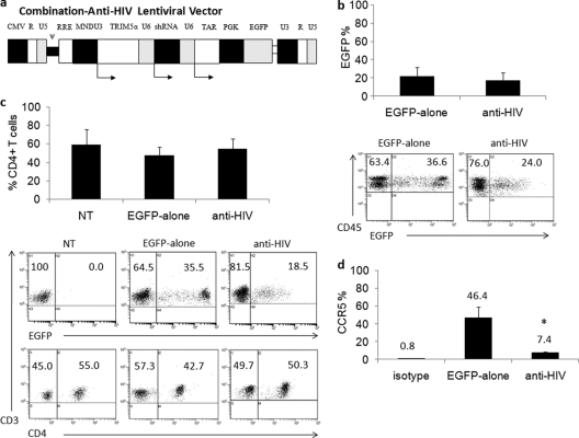
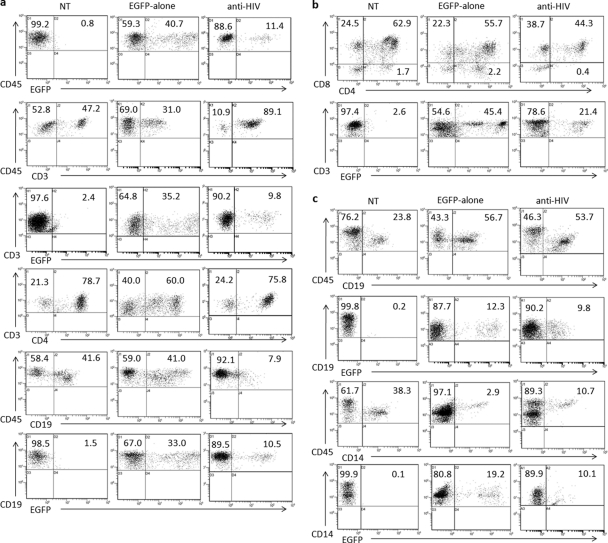
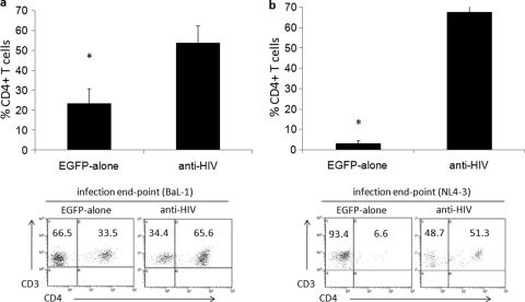
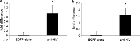
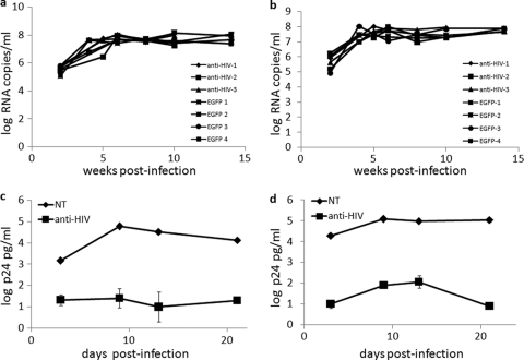
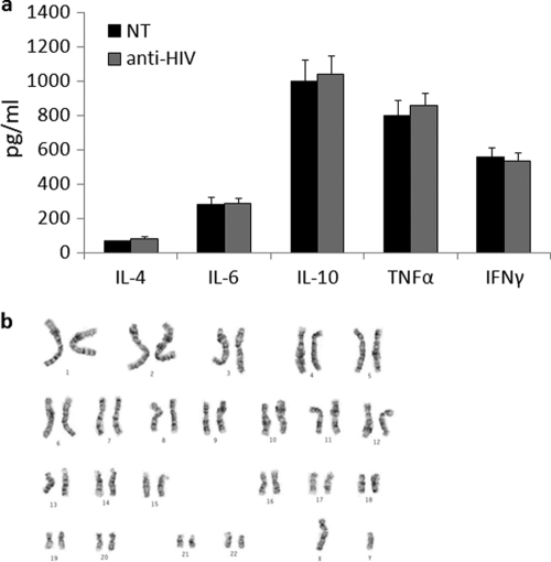
HIV gene therapy has the potential to offer an alternative to the use of current small-molecule antiretroviral drugs as a treatment strategy for HIV-infected individuals. Therapies designed to administer HIV-resistant stem cells to an infected patient may also provide a functional cure, as observed in a bone marrow transplant performed with hematopoietic stem cells (HSCs) homozygous for the CCR5-Δ32-bp allele. In our current studies, preclinical evaluation of a combination anti-HIV lentiviral vector was performed, in vivo, in humanized NOD-RAG1(-/-) IL2rγ(-/-) knockout mice. This combination vector, which displays strong preintegration inhibition of HIV-1 infection in vitro, contains a human/rhesus macaque TRIM5α isoform, a CCR5 short hairpin RNA (shRNA), and a TAR decoy. Multilineage hematopoiesis from anti-HIV lentiviral vector-transduced human CD34(+) HSCs was observed in the peripheral blood and in various lymphoid organs, including the thymus, spleen, and bone marrow, of engrafted mice. Anti-HIV vector-transduced CD34(+) cells displayed normal development of immune cells, including T cells, B cells, and macrophages. The anti-HIV vector-transduced cells also displayed knockdown of cell surface CCR5 due to the expression of the CCR5 shRNA. After in vivo challenge with either an R5-tropic BaL-1 or X4-tropic NL4-3 strain of HIV-1, maintenance of human CD4(+) cell levels and a selective survival advantage of anti-HIV gene-modified cells were observed in engrafted mice. The data provided from our study confirm the safety and efficacy of this combination anti-HIV lentiviral vector in a hematopoietic stem cell gene therapy setting for HIV and validates its potential application in future clinical trials.


Figures

Fig 1
Fig 1
Combination anti-HIV lentiviral vector, peripheral blood engraftment of transduced cells, and CCR5 downregulation. A third-generation lentiviral vector, CCLc-x-PGK-EGFP, was utilized to generate the combination anti-HIV construct. (a) A human/rhesus macaque TRIM5α isoform was driven under the control of the MNDU3 promoter, and a CCR5 shRNA and a TAR decoy were driven under separate human polymerase III U6 small RNA promoters. These three anti-HIV genes were inserted upstream from the EGFP reporter gene. (b) CD34+ HSCs were transduced with the control EGFP-alone vector or the anti-HIV vector and transplanted into NRG pups. Transplanted mice were screened for human CD45 and EGFP expression in the peripheral blood for engraftment of transduced cells, either EGFP-alone (n = 12) or anti-HIV (n = 14). (c) The peripheral blood of nontransduced (NT) (n = 14), EGFP-alone (n = 12), and anti-HIV (n = 14) cell-engrafted mice was analyzed for human T cells with antibodies specific for CD3 and CD4 and also for expression of EGFP. (d) CD4+ human splenocytes were analyzed for the expression of CCR5 (n = 5 for EGFP-alone and n = 11 for anti-HIV). Bar graphs display averages and standard deviations for each cohort. Statistical significance (P < 0.05) is represented by an asterisk.
Fig 2
Fig 2
Engraftment of lymphoid organs in transplanted NRG mice. NRG mice were transplanted with CD34+ HSCs that were either nontransduced (NT) or transduced with a control EGFP-alone vector or the anti-HIV lentiviral vector. Upon engraftment, various lymphoid organs, including the spleen (a), thymus (b), and bone marrow (c), were analyzed for human cell engraftment. Flow cytometry was performed to detect EGFP expression along with total human leukocytes (CD45), T cells (CD3, CD4, and CD8), B cells (CD19), and macrophages (CD14). Data are representative of mice for each cohort. Complete data sets for each cohort of mice are included in Table 2.
Fig 3
Fig 3
Detection of human CD4+ T cells in the peripheral blood of HIV-1-infected NRG humanized mice. (a and b) NRG mice successfully engrafted with either control EGFP-alone or combination anti-HIV vector-transduced cells were infected i.v. with either an R5-tropic BaL-1 (a) or an X4-tropic NL4-3 (b) strain of HIV-1. At various weeks postinfection, mice were bled and analyzed by FACS for total human CD4+ cell percentage. Solid lines represent anti-HIV cell-engrafted mice. Dashed lines represent control EGFP-alone cell-engrafted mice. (c and d) Comparisons between CD4+ T cell level averages preinfection and postinfection were performed for both the BaL-1 (c)- and NL4-3 (d)-infected mice engrafted with either EGFP-alone or anti-HIV vector-transduced cells. Bar graphs display averages and standard deviations from four mice for each cohort for each set of infections. Statistical significance (P < 0.05) is represented by an asterisk. Representative flow cytometry plots are displayed.
Fig 4
Fig 4
Detection of human CD4+ T cells in the spleens of HIV-1-infected NRG humanized mice. NRG mice successfully engrafted with either control EGFP-alone or combination anti-HIV vector-transduced cells were infected i.v. with either an R5-tropic BaL-1 (a) or an X4-tropic NL4-3 (b) strain of HIV-1. After completion of the in vivo challenge experiments, infected mice were sacrificed and the spleens were analyzed by flow cytometry for CD4+ T cell (CD3+) levels. Bar graphs display averages and standard deviations from four mice for each cohort for each infection. Statistical significance (P < 0.05) is represented by an asterisk. Representative flow cytometry plots are displayed.
Fig 5
Fig 5
Selective survival advantage of anti-HIV gene-modified cells in HIV-1-infected NRG mice. Mice successfully engrafted with either control EGFP-alone or combination anti-HIV vector-transduced cells were infected i.v. with either an R5-tropic BaL-1 (a) or an X4-tropic NL4-3 (b) strain of HIV-1. At various weeks postinfection, mice were bled and analyzed by FACS for EGFP+/CD4+ human cell percentage. Fold difference in EGFP+/CD4+ T cell level averages preinfection and postinfection are displayed for both the EGFP-alone and anti-HIV vector-transduced cells. Bar graphs display averages and standard deviations from four mice for each cohort for each set of infections. Statistical significance (P < 0.05) is represented by an asterisk.
Fig 6
Fig 6
Detection of in vivo plasma viremia and in vitro HIV-1 challenge of sorted spleen T cells. (a and b) NRG mice successfully engrafted with either control EGFP-alone or combination anti-HIV vector-transduced cells were infected i.v. with either an R5-tropic BaL-1 (a) or an X4-tropic NL4-3 (b) strain of HIV-1. At various weeks postinfection, mice were bled and the plasma was analyzed by Q-PCR using a primer/probe pair specific for the HIV pol gene. (c and d) In vitro HIV-1 challenge experiments were performed on human CD3+ T cells, both nontransduced (EGFP) and anti-HIV vector transduced (EGFP+), with an R5-tropic BaL-1 (c) or an X4-tropic NL4-3 (d) strain of HIV-1. At various days postinfection, culture supernatants were collected and analyzed for p24 by antigen ELISA. p24 ELISAs were performed in triplicate.
Fig 7
Fig 7
Cytokine expression and karyotypic analysis of anti-HIV vector-transduced cells. (a) Anti-HIV gene-modified T cells from spleen were sorted based on EGFP/CD3 expression and stimulated with IL-2 and PHA. At day 3 poststimulation, culture supernatants were analyzed by FACS for expression of IL-4, IL-6, IL-10, TNF-α, and IFN-γ. Cytokine expression experiments were performed in triplicate. (b) A representative karyotyping analysis of anti-HIV vector-transduced human CD34+ HSCs. Karyotypic analyses were performed in duplicate.

Similar articles

Cited by

References

    1. An DS, et al. 2007. Stable reduction of CCR5 by RNAi through hematopoietic stem cell transplant in non-human primates. Proc. Natl. Acad. Sci. U. S. A. 104:13110–13115 - PMC - PubMed
    1. Anderson J, et al. 2007. Safety and efficacy of a lentiviral vector containing three anti-HIV genes—CCR5 ribozyme, tat-rev siRNA, and TAR decoy—in SCID-hu mouse-derived T cells. Mol. Ther. 15:1182–1188 - PubMed
    1. Anderson J, Akkina R. 2007. Complete knockdown of CCR5 by lentiviral vector-expressed siRNAs and protection of transgenic macrophages against HIV-1 infection. Gene Ther. 14:1287–1297 - PubMed
    1. Anderson J, Akkina R. 2008. Human immunodeficiency virus type 1 restriction by human-rhesus chimeric tripartite motif 5alpha (TRIM 5alpha) in CD34+(+) cell-derived macrophages in vitro and in T cells in vivo in severe combined immunodeficient (SCID-hu) mice transplanted with human fetal tissue. Hum. Gene Ther. 19:217–228 - PubMed
    1. Anderson JS, Javien J, Nolta JA, Bauer G. 2009. Preintegration HIV-1 inhibition by a combination lentiviral vector containing a chimeric TRIM5alpha protein, a CCR5 shRNA, and a TAR decoy. Mol. Ther. 17:2103–2114 - PMC - PubMed

Publication types