. 2012 Mar 7;483(7389):336-40.
doi: 10.1038/nature10879.

Phase transitions in the assembly of multivalent signalling proteins

Affiliations

Phase transitions in the assembly of multivalent signalling proteins

Pilong Li et al. Nature. .

Abstract

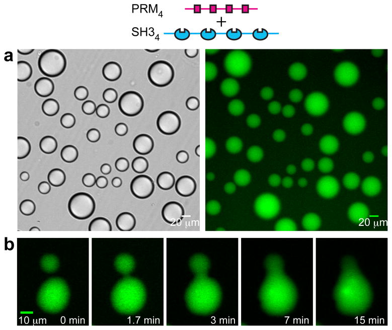
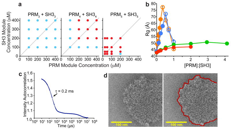
Cells are organized on length scales ranging from ångström to micrometres. However, the mechanisms by which ångström-scale molecular properties are translated to micrometre-scale macroscopic properties are not well understood. Here we show that interactions between diverse synthetic, multivalent macromolecules (including multi-domain proteins and RNA) produce sharp liquid-liquid-demixing phase separations, generating micrometre-sized liquid droplets in aqueous solution. This macroscopic transition corresponds to a molecular transition between small complexes and large, dynamic supramolecular polymers. The concentrations needed for phase transition are directly related to the valency of the interacting species. In the case of the actin-regulatory protein called neural Wiskott-Aldrich syndrome protein (N-WASP) interacting with its established biological partners NCK and phosphorylated nephrin, the phase transition corresponds to a sharp increase in activity towards an actin nucleation factor, the Arp2/3 complex. The transition is governed by the degree of phosphorylation of nephrin, explaining how this property of the system can be controlled to regulatory effect by kinases. The widespread occurrence of multivalent systems suggests that phase transitions may be used to spatially organize and biochemically regulate information throughout biology.


Figures

Figure 1
Figure 1. Macroscopic and microscopic phase transitions in multivalent SH3+PRM systems
a, Liquid droplets observed by differential interference contrast microscopy (left) and widefield fluorescence microscopy (right) when 300 μM SH34, 300 μM PRM4 (module concentrations; molecule concentrations are 75 μM) and 0.5 μM OG-SH34 are mixed. b, Time-lapse imaging of merging droplets formed as in a.
Figure 2
Figure 2. Multivalency drives phase separation and likely a sol-gel transition in the droplet phase
a, Phase diagrams of multivalent SH3 and PRM proteins. Concentrations are in terms of modules. Red and blue circles indicate phase separation and no phase separation, respectively. b. Rg values determined from SAXS data collected during titrations of PRM proteins into SH35. Closed circles indicate the absence of phase separation; open circles indicate data collected on supernatant phase, separated from droplets by centrifugation. Titrations used PRM4 (orange), PRM2 (blue), PRM1 (green), PRM(H)1 (red). Error bars represent standard deviation calculated from 5- 10 independent measurements of I versus Q. c. Intensity autocorrelation curve of light scattered at 90 ° from the pooled droplet phase of SH35 + PRM(N-WASP)8. d. Electron cryo-microscopic image of a droplet formed by SH35 + PRM5. Right panel shows edge of the structure with a red line.
Figure 3
Figure 3. Co-expression of SH35 and PRM5 in cells produces dynamic puncta
a, From left to right, panels show mCherry-SH35, eGFP-PRM5 and overlay in a cell expressing both proteins. Note that non-uniform eGFP fluorescence in the puncta is due to mCherry-eGFP FRET rather than differential localization of the proteins (Supplementary Fig. 17). b, Both mCherry and eGFP fluorescence recover rapidly after photobleaching.
Figure 4
Figure 4. Phase transition correlates to biochemical activity transition in the nephrin/Nck/N-WASP system
a, Interactions of nephrin, Nck and N-WASP. b-d, Phase diagrams of N-WASP and Nck alone (b) or in the presence of 4.5 μm doubly- (c) or 3 μm triply-phosphorylated (d) nephrin tail peptides. Red and blue circles same as in Figure 2a. e, Half-time (t1/2) of N-WASP-stimulated actin assembly by the Arp2/3 complex as a function of N-WASPΔ concentration. Vertical dashed line indicates the phase separation boundary determined in separate assays without actin and Arp2/3 complex. f, Rhodamine-actin (10 % labeled, 4 μM), 300 nM Alexa 488–phalloidin and 10 nM Arp2/3 complex were added to droplets containing triply-phosphorylated nephrin, Nck and N-WASP and imaged by confocal microscopy. Top panel = rhodamine; bottom panel = Alexa 488. Scale bar = 10 μm.

References

    1. Jones N, et al. Nck adaptor proteins link nephrin to the actin cytoskeleton of kidney podocytes. Nature. 2006;440:818–823. - PubMed
    1. Flory PJ. Principles of Polymer Chemistry. Cornell University Press; 1953.
    1. Cohen RJ, Benedek GB. Equilibrium and Kinetic Theory of Polymerization and the Sol-Gel Transition. J Phys Chem. 1982;86:3696–3714.
    1. Lehn JM. Supramolecular Polymer Chemistry--Scope and Perspectives. Polym Int. 2002;51:825–839.
    1. Tanaka F. Polymer Physics: Applications to Molecular Association and Thermoreversible Gelation. Cambridge University Press; 2011.

Publication types

MeSH terms