

Quantification of myocardial blood flow with 82Rb positron emission tomography: clinical validation with 15O-water
- PMID: 22398957
- PMCID: PMC3342496
- DOI: 10.1007/s00259-012-2082-3
Quantification of myocardial blood flow with 82Rb positron emission tomography: clinical validation with 15O-water
Abstract
Purpose: Quantification of myocardial blood flow (MBF) with generator-produced (82)Rb is an attractive alternative for centres without an on-site cyclotron. Our aim was to validate (82)Rb-measured MBF in relation to that measured using (15)O-water, as a tracer 100% of which can be extracted from the circulation even at high flow rates, in healthy control subject and patients with mild coronary artery disease (CAD).
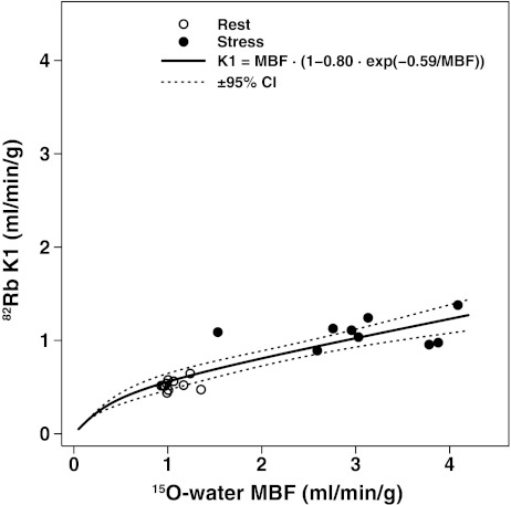
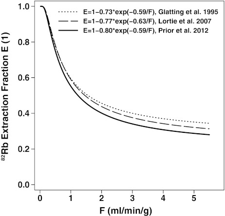
Methods: MBF was measured at rest and during adenosine-induced hyperaemia with (82)Rb and (15)O-water PET in 33 participants (22 control subjects, aged 30 ± 13 years; 11 CAD patients without transmural infarction, aged 60 ± 13 years). A one-tissue compartment (82)Rb model with ventricular spillover correction was used. The (82)Rb flow-dependent extraction rate was derived from (15)O-water measurements in a subset of 11 control subjects. Myocardial flow reserve (MFR) was defined as the hyperaemic/rest MBF. Pearson's correlation r, Bland-Altman 95% limits of agreement (LoA), and Lin's concordance correlation ρ (c) (measuring both precision and accuracy) were used.
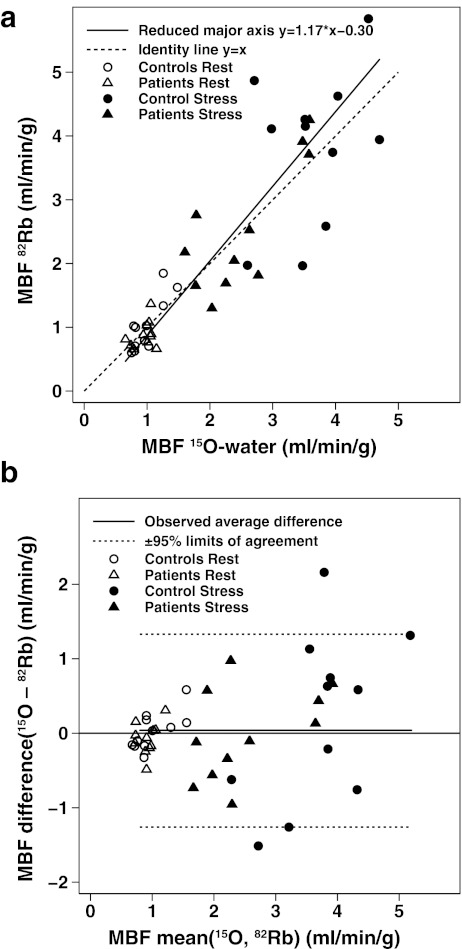
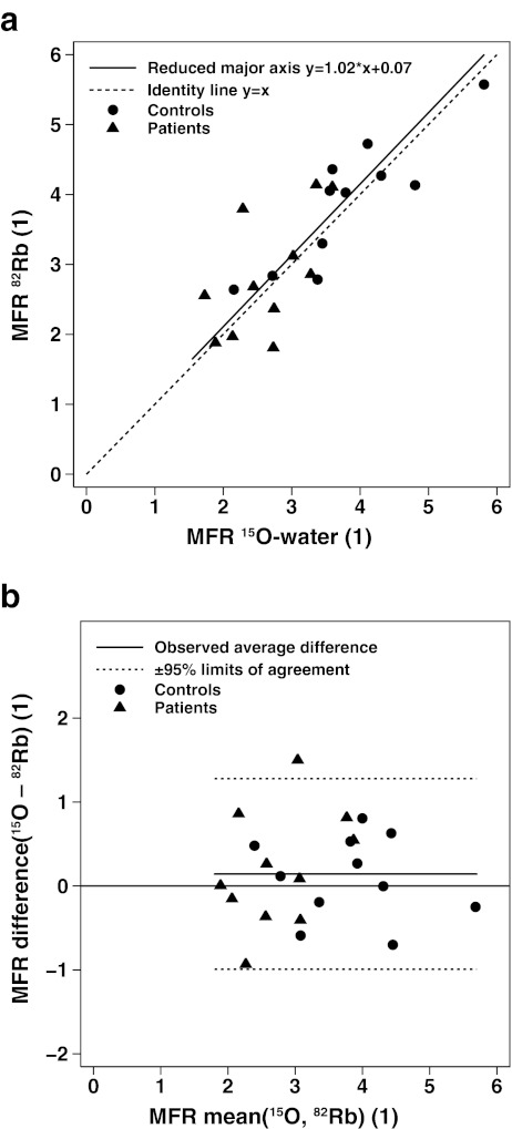
Results: Over the entire MBF range (0.66-4.7 ml/min/g), concordance was excellent for MBF (r = 0.90, [(82)Rb-(15)O-water] mean difference ± SD = 0.04 ± 0.66 ml/min/g, LoA = -1.26 to 1.33 ml/min/g, ρ(c) = 0.88) and MFR (range 1.79-5.81, r = 0.83, mean difference = 0.14 ± 0.58, LoA = -0.99 to 1.28, ρ(c) = 0.82). Hyperaemic MBF was reduced in CAD patients compared with the subset of 11 control subjects (2.53 ± 0.74 vs. 3.62 ± 0.68 ml/min/g, p = 0.002, for (15)O-water; 2.53 ± 1.01 vs. 3.82 ± 1.21 ml/min/g, p = 0.013, for (82)Rb) and this was paralleled by a lower MFR (2.65 ± 0.62 vs. 3.79 ± 0.98, p = 0.004, for (15)O-water; 2.85 ± 0.91 vs. 3.88 ± 0.91, p = 0.012, for (82)Rb). Myocardial perfusion was homogeneous in 1,114 of 1,122 segments (99.3%) and there were no differences in MBF among the coronary artery territories (p > 0.31).
Conclusion: Quantification of MBF with (82)Rb with a newly derived correction for the nonlinear extraction function was validated against MBF measured using (15)O-water in control subjects and patients with mild CAD, where it was found to be accurate at high flow rates. (82)Rb-derived MBF estimates seem robust for clinical research, advancing a step further towards its implementation in clinical routine.
Figures
References
-
- Gould KL, Goldstein RA, Mullani NA, Kirkeeide RL, Wong WH, Tewson TJ, et al. Noninvasive assessment of coronary stenoses by myocardial perfusion imaging during pharmacologic coronary vasodilation. VIII. Clinical feasibility of positron cardiac imaging without a cyclotron using generator-produced rubidium-82. J Am Coll Cardiol. 1986;7:775–789. doi: 10.1016/S0735-1097(86)80336-9. - DOI - PubMed
-
- Sampson UK, Dorbala S, Limaye A, Kwong R, Di Carli MF. Diagnostic accuracy of rubidium-82 myocardial perfusion imaging with hybrid positron emission tomography/computed tomography in the detection of coronary artery disease. J Am Coll Cardiol. 2007;49:1052–1058. doi: 10.1016/j.jacc.2006.12.015. - DOI - PubMed
Publication types
MeSH terms
Substances
LinkOut - more resources
Full Text Sources
Miscellaneous
