

Consequences of fuzziness in the NFκB/IκBα interaction
- PMID: 22399319
- PMCID: PMC3603378
- DOI: 10.1007/978-1-4614-0659-4_5
Consequences of fuzziness in the NFκB/IκBα interaction
Abstract
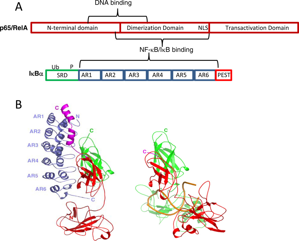
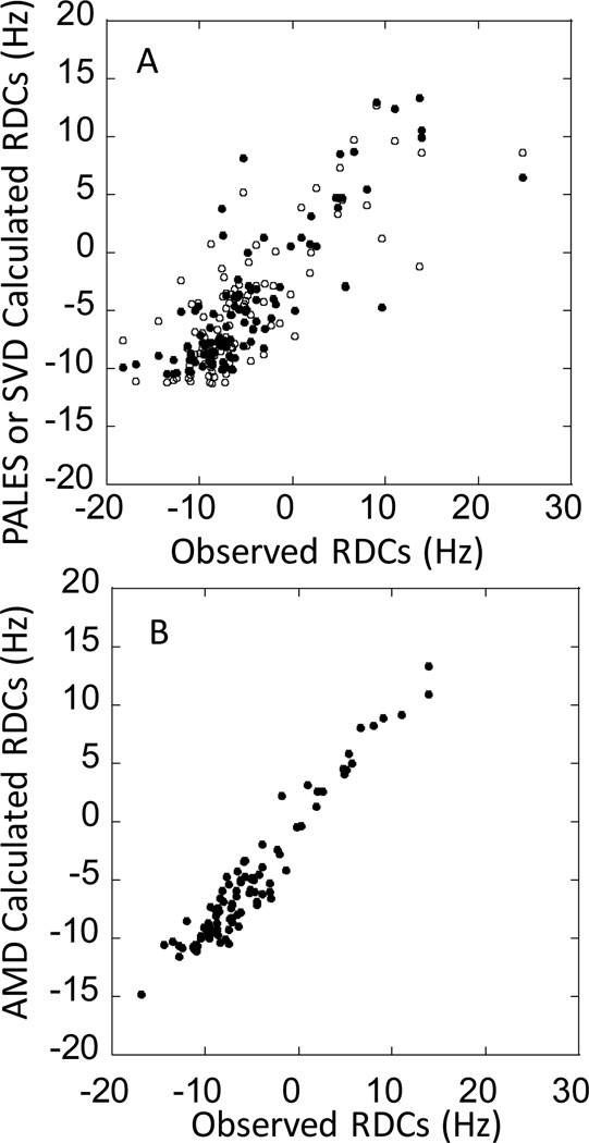
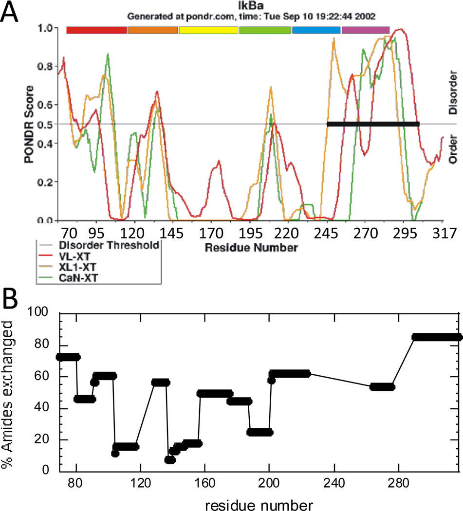
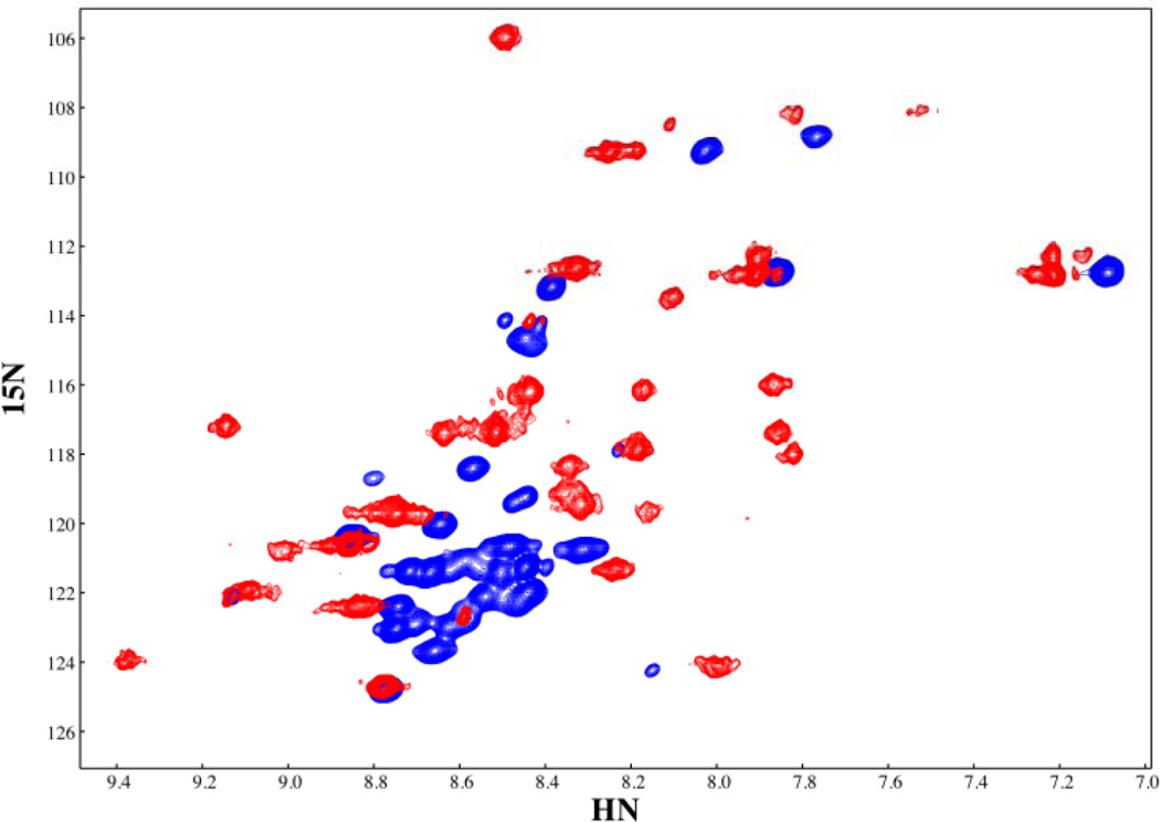
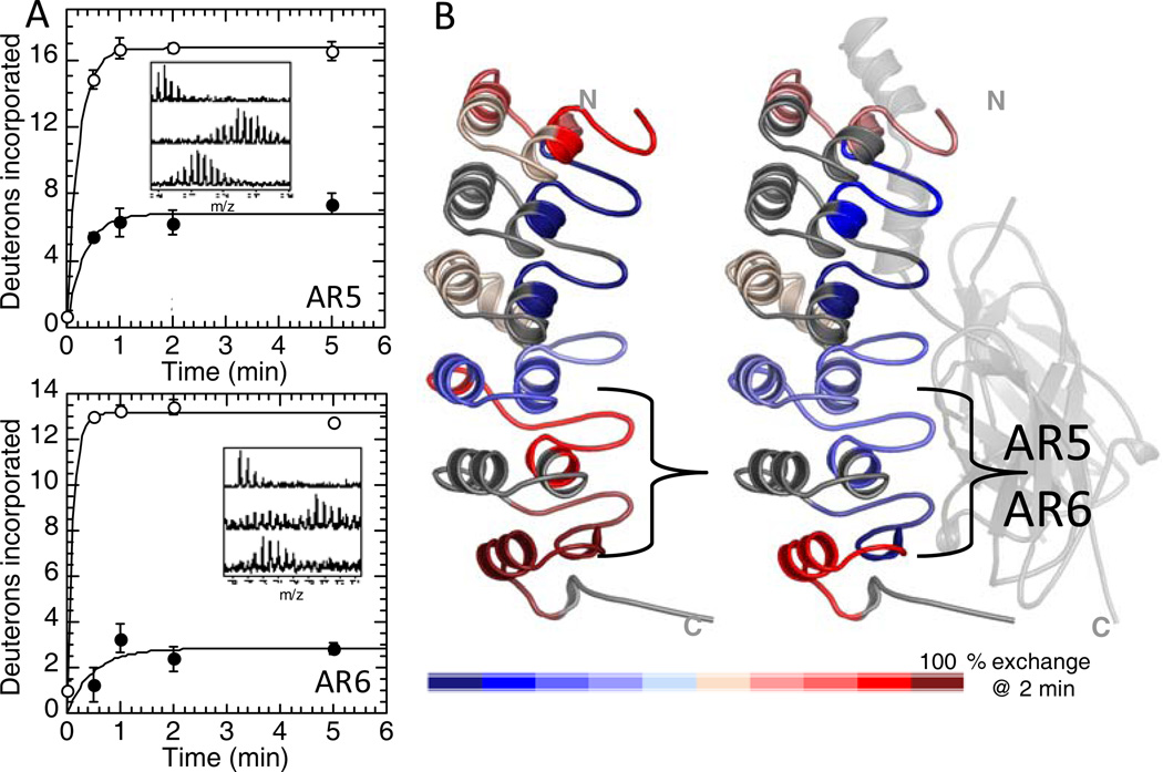
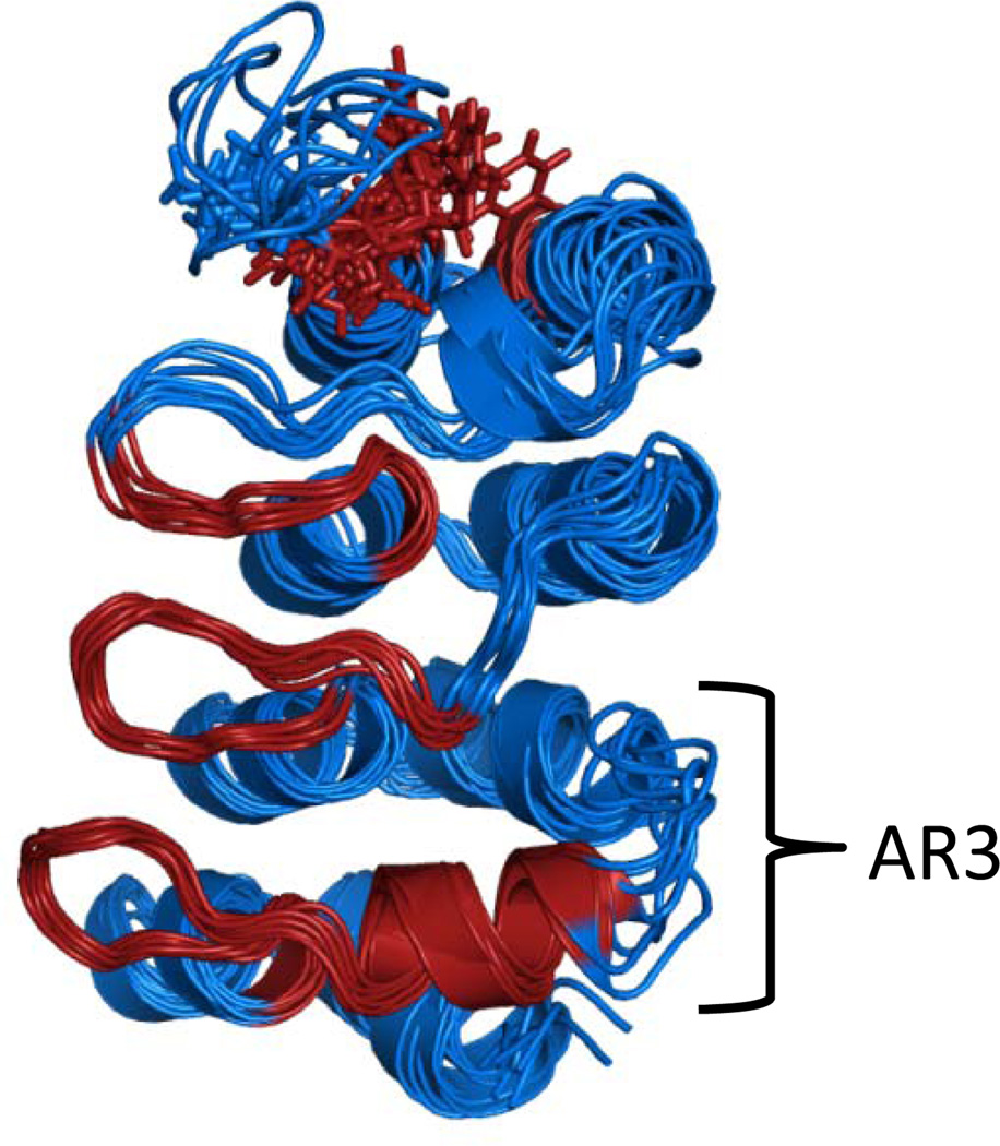
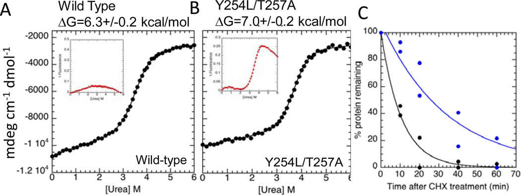
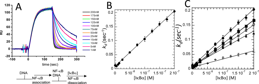
This chapter provides a short review of various biophysical experiments that have been applied to the inhibitor of kappa B, IκBα and its binding partner, nuclear factor kappa B, or NFκB. The picture that emerges from amide hydrogen/deuterium exchange, NMR and binding kinetics experiments is one in which parts of both proteins are "fuzzy" in the free-state and some parts remain "fuzzy" in the NFκB-IκBα complex. The NFκB family of transcription factors responds to inflammatory cytokines with rapid transcriptional activation, in which NFκB enters the nucleus and binds DNA. Just as rapidly as transcription is activated, it is subsequently repressed by newly synthesized IκBα?that also enters the nucleus and removes NFκB from the DNA. Because IκBα?is an ankyrin repeat protein, it's "fuzziness" can be controlled by mutagenesis to stabilized the folded state. Experimental comparison with such stabilized mutants helps provide evidence that much of the system control depends on the "fuzziness" of IκBα.
Figures
Similar articles
-
Detection of a ternary complex of NF-kappaB and IkappaBalpha with DNA provides insights into how IkappaBalpha removes NF-kappaB from transcription sites.Proc Natl Acad Sci U S A. 2011 Jan 25;108(4):1367-72. doi: 10.1073/pnas.1014323108. Epub 2011 Jan 10. Proc Natl Acad Sci U S A. 2011. PMID: 21220295 Free PMC article.
-
Regions of IkappaBalpha that are critical for its inhibition of NF-kappaB.DNA interaction fold upon binding to NF-kappaB.Proc Natl Acad Sci U S A. 2006 Dec 12;103(50):18951-6. doi: 10.1073/pnas.0605794103. Epub 2006 Dec 5. Proc Natl Acad Sci U S A. 2006. PMID: 17148610 Free PMC article.
-
Visualization of the nanospring dynamics of the IkappaBalpha ankyrin repeat domain in real time.Proc Natl Acad Sci U S A. 2011 Jun 21;108(25):10178-83. doi: 10.1073/pnas.1102226108. Epub 2011 May 31. Proc Natl Acad Sci U S A. 2011. PMID: 21628581 Free PMC article.
-
Molecular mechanisms of system control of NF-kappaB signaling by IkappaBalpha.Biochemistry. 2010 Mar 2;49(8):1560-7. doi: 10.1021/bi901948j. Biochemistry. 2010. PMID: 20055496 Free PMC article. Review.
-
Role of disorder in IκB-NFκB interaction.IUBMB Life. 2012 Jun;64(6):499-505. doi: 10.1002/iub.1044. Epub 2012 May 9. IUBMB Life. 2012. PMID: 22573609 Free PMC article. Review.
Cited by
-
Post-translational Modifications of IκBα: The State of the Art.Front Cell Dev Biol. 2020 Nov 5;8:574706. doi: 10.3389/fcell.2020.574706. eCollection 2020. Front Cell Dev Biol. 2020. PMID: 33224945 Free PMC article. Review.
-
Regulation and function of nuclear IκBα in inflammation and cancer.Am J Clin Exp Immunol. 2012 May 25;1(1):56-66. Print 2012. Am J Clin Exp Immunol. 2012. PMID: 23885315 Free PMC article.
-
Histone demethylase KDM5B licenses macrophage-mediated inflammatory responses by repressing Nfkbia transcription.Cell Death Differ. 2023 May;30(5):1279-1292. doi: 10.1038/s41418-023-01136-x. Epub 2023 Mar 13. Cell Death Differ. 2023. PMID: 36914768 Free PMC article.
References
-
- Hoffmann A, Levchenko A, Scott ML, Baltimore D. The IkappaB-NF-kappaB signaling module: temporal control and selective gene activation. Science. 2002;298:1241–1245. - PubMed
-
- Hoffmann A, Baltimore D. Circuitry of nuclear factor kappaB signaling. Immunol Rev. 2006;210:171–186. - PubMed
-
- Ghosh S, May MJ, Kopp EB. NF-kappa B and Rel proteins: evolutionarily conserved mediators of immune responses. Annu. Rev. Immunol. 1998;16:225–260. - PubMed
-
- Kumar A, Takada Y, Boriek AM, Aggarwal BB. Nuclear factor-kappaB: its role in health and disease. J. Mol. Med. 2004;82:434–448. - PubMed
-
- Chen FE, Huang DB, Chen YQ, Ghosh G. Crystal structure of p50/p65 heterodimer of transcription factor NF-kappaB bound to DNA. Nature. 1998;391:410–413. - PubMed
MeSH terms
Substances
Grants and funding
LinkOut - more resources
Full Text Sources
