. 2012:6:11-21.
doi: 10.4137/BBI.S8451. Epub 2012 Feb 1.

Structural and functional insights on the Myosin superfamily

Affiliations

Structural and functional insights on the Myosin superfamily

Divya P Syamaladevi et al. Bioinform Biol Insights. 2012.

Abstract

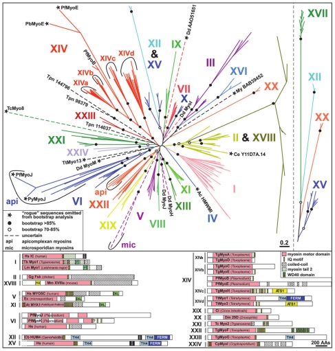
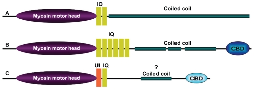
The myosin superfamily is a versatile group of molecular motors involved in the transport of specific biomolecules, vesicles and organelles in eukaryotic cells. The processivity of myosins along an actin filament and transport of intracellular 'cargo' are achieved by generating physical force from chemical energy of ATP followed by appropriate conformational changes. The typical myosin has a head domain, which harbors an ATP binding site, an actin binding site, and a light-chain bound 'lever arm', followed often by a coiled coil domain and a cargo binding domain. Evolution of myosins started at the point of evolution of eukaryotes, S. cerevisiae being the simplest one known to contain these molecular motors. The coiled coil domain of the myosin classes II, V and VI in whole genomes of several model organisms display differences in the length and the strength of interactions at the coiled coil interface. Myosin II sequences have long-length coiled coil regions that are predicted to have a highly stable dimeric interface. These are interrupted, however, by regions that are predicted to be unstable, indicating possibilities of alternate conformations, associations to make thick filaments, and interactions with other molecules. Myosin V sequences retain intermittent regions of strong and weak interactions, whereas myosin VI sequences are relatively devoid of strong coiled coil motifs. Structural deviations at coiled coil regions could be important for carrying out normal biological function of these proteins.

Keywords: coiled coil; myosin domain architecture; myosin structure.


Figures

Figure 1
Figure 1
Different classes of myosin superfamily: Phylogenetic tree of 114 myosin sequences showing 24 classes of myosins (Foth et al; Copyright (2006) National Academy of Sciences, USA.).
Figure 2
Figure 2
Domain architecture. The overall length and domain organization vary considerably between the subclass members of myosin superfamily. Domain architectures of human (A) myosin II (NP_005954.3: 1939 amino acids) (B) myosin Va (NP_000250.3:1855 amino acids) and (C) myosin VI (NP_004990.3: 1285 amino acids). The length of predicted coiled coil domain using COILS program reflects the variation in length and architecture of the coiled coil domains. Lengths of coiled coil of human (A) myosin II (NP_005954.3 : residue 843–1931), (B) myosin Va (NP_000250.3: residue 911–1104; residue 1153–1234; residue 1339–1445) and (C) myosin VI (NP_004990.3: residue 864–1030) are shown in green.
Figure 3
Figure 3
Gene organization of myosin superfamily. Myosin genes are highly interrupted and contain large number of exons. (A) human myosin II (NP_005954.3: 40 exons; Transcript length: 6,023 bps), (B) human myosin Va (NP_000250.3: 41 exons; Transcript length: 12,225 bps) and (C) human myosin VI (NP_004990.3: 35 exons; Transcript length: 8,662 bps).
Figure 4
Figure 4
Crystal structures of Cargo Binding Domain (CBD). (A) crystal structure of Myosin V CBD from Saccharomyces cerevisiae (PDB ID: 2F6H), (B) NMR structure of Myosin VI CBD from Mus musculus (PDB ID: 2KIA).
Figure 5
Figure 5
Coiled coils interaction strength analysis protocol. Molecular models of myosin coiled coils were made using tropomyosin as template. The models were generated using MODELLER program. The long models were split into penta-heptad sized fragments and interaction energy values were calculated using COILCHECK program.
Figure 6
Figure 6
COILCHECK based inter-protomer interactions. (A) Left: Pictorial representation of parallel coiled coil crystal structures depicting the interaction energy. Each block corresponds to a 35 residue window. Green blocks are regions of low inter-protomer interaction energy in COILCHECK calculations. Right top: PyMOL representation of coiled coil of BZIP transcription factor bound to DNA (PDB ID: 1IO4). Vdw1, vdw2 are the van der Waals energies for the first and second penta-heptad fragments of the coiled coil respectively. Van der Waals contribution in the first half that interacts with DNA is very less compared to the second half. The total normalized energy per residue for this region is also positive (shown on Right bottom). Right bottom: Pictorial representation of parallel coiled coils complexed with DNA. Red block represents the high calculated interaction energy that indicates the absence purely dimeric coiled coil interaction. This high interaction energy reflects either a poorly interacting dimer, a higher order assembly or interaction with other proteins. (B) Region II and III of GEF domain of sec2p (shown in green) has low COILCHECK calculated interaction energies as compared to the N terminal segment (shown in green) that is not a coiled coil and the C terminal segment (shown in red) that is weakly interacting. (C) Normalized energy for 14 residue long continuous fragments of tropomyosin (1C1G) structure. The positive energy regions (circled in red) correspond either to alanin repeats or breaks in hydrophobic cores. Alanine repeats and sequence periods are quasi-equivalent to actin binding sites. (D) ΔE is the difference in normalized energy values of wild type GCN4 and single point in silico mutants. A single residue was mutated at a time in a,d, e and g positions of an ideal heptad of GCN4. When positions a and d (Valine and Leucine) were mutated to hydrophobic residues the mutants showed least ΔE (shown in light green) and when mutated to charged residues ΔE was higher and destabilizing (shown in orange). Position g when mutated to any other residue did not show much deviation (light green). Position e when mutated to any other residue we found a stabilizing effect (dark green). The color scale from red to green is shown on the left side of the graph. The numbers (in the negative side) corresponding to each bar in the graph are the normalized stabilization energy for that fragment. The two lines of numbers (in the positive side) corresponds to the total electrostatic (first line from top) and Van der Waals energies in kJ/mol (second line from top).
Figure 7
Figure 7
Inter-protomer interaction strength. Non-uniform inter-protomer interaction strength in predicted coiled coil regions are color coded and represented as boxes. Each box corresponds to 35 amino acids. Strongly interacting regions are shown in green and non interacting regions are shown in red. An intermediate level of interaction is also possible and shown in lighter shade of red. (A) human myosinII NP_001070654.1. (B) human myosin Va NP_000250.3. (C) human myosin VI (NP_004990.3). (D) Human myosin VI normalized energy per residue plotted against the pentaheptads. The first line of numbers in the positive side of the graph corresponds to the total electrostatic interaction energy and the second line corresponds to the total Van der Waals interaction energy.

References

    1. Richards TA, Cavalier-Smith T. Myosin domain evolution and the primary divergence of eukayotes. Nature. 2005;436:1113–8. - PubMed
    1. Goodson HV, Spudich JA. Molecular evolution of myosin family:relationships derived from comparisons of amino acid sequences. Proc Natl Acad Sci U S A. 1993;90:659–3. - PMC - PubMed
    1. De Lozanne A, Spudich JA. Disruption of the Dictyostelium myosin heavy chain gene by homologous recombination. Science. 1987;236:1086–91. - PubMed
    1. Knecht DA, Loomis WF. Antisense RNA inactivation of myosin heavy chain gene expression in Dictyostelium discoideum. Science. 1987;236:1081–5. - PubMed
    1. Tuxworth RI, Titus MA. Unconventional myosins: anchors in the membrane traffic relay. Traffic. 2000;1:11–8. - PubMed

LinkOut - more resources