

Pulmonary large cell neuroendocrine carcinoma diagnosed in a brain metastasis
- PMID: 22400077
- PMCID: PMC3294230
Pulmonary large cell neuroendocrine carcinoma diagnosed in a brain metastasis
Abstract
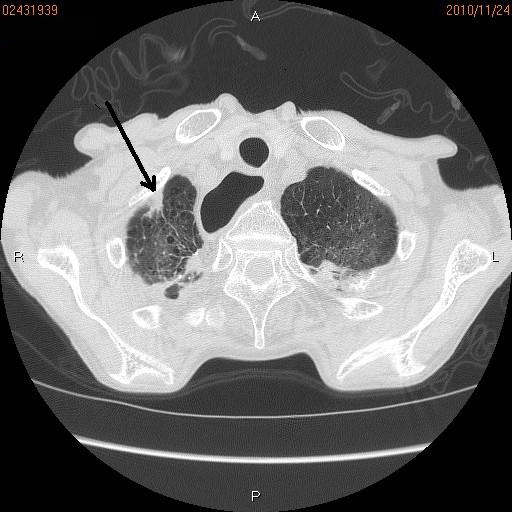
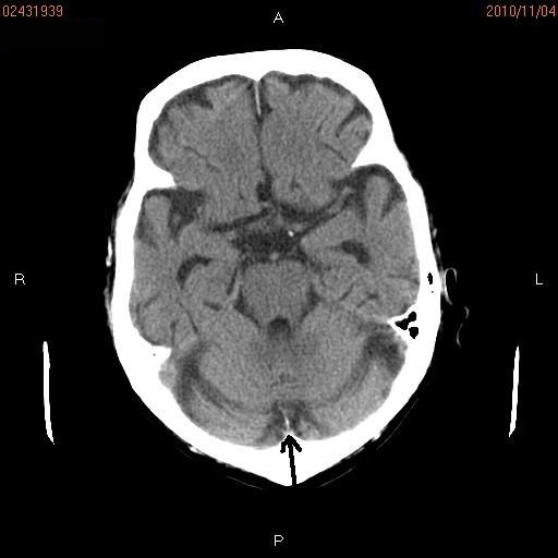
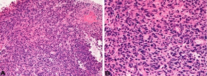
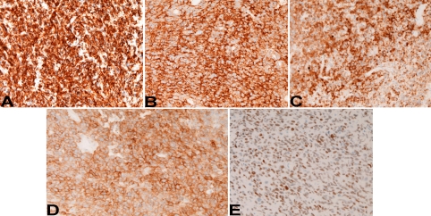
The author herein reports a large cell neuroendocrine carcinoma (LCNEC) of the lung diagnosed at a brain metastasis without clinical data. A 70-year-old man underwent esophagectomy for esophageal squamous cell carcinoma, and was treated with chemotherapy. At 72 years of age, he was found to have prostatic well differentiated adenocarcinoma, and treated by estrogen. At 78 years of age, he was pointed out to have gastric advanced tumor, and the biopsy showed moderately differentiated tubular adenocarcinoma. The gastric carcinoma was treated by chemotherapy. At 79 years of age, he was shown to have right lung shadow (2 cm in diameter) and brain shadow (cerebellar vermis) of 1 cm in diameter. Multiple biopsy and cytology of the lung failed to detect carcinoma cells. Biopsy of the brain was performed. The biopsy showed medullary undifferentiated carcinoma. Immunohistochemically, the tumor cells were positive for pancytokeratin AE1/3, synaptophysin, CD56 (NCAM), p53, Ki67 (labeling 40%), KIT and TTF-1, but were negative for vimentin, chromogranin, neuron-specific enolase and PDGFRA. A pathological diagnosis of metastatic LCNEC form the lung was made. A molecular genetic analysis for KIT (exons 9, 11, 13, and 17) and PDGFRA (exons 12 and 18) genes identified no mutations of the KIT and PDGFRA genes. The patients died of carcinomatosis one month after the diagnosis. In conclusion, careful histological and immunohistochemical examination can diagnose LCNEC of the lung at the metastatic site.
Keywords: LCNEC; brain; immunohistochemistry; lung; metastasis.
Figures
References
-
- Terada T, Kawaguchi M, Furukawa K, Sekido Y, Osamura Y. Minute mixed ductalendocrine 10|carcinoma of the pancreas with predominant intraductal growth. Pathol Int. 2002;52:740–746. - PubMed
-
- Terada T, Tanigichi M. Intraductal oncocytic papillary neoplasm of the liver. Pathol Int. 2004;54:116–123. - PubMed
-
- Terada T, Kawaguchi M. Primary clear cell adenocarcinoma of the peritoneum. Tohoku J Exp Med. 2005;206:271–275. - PubMed
-
- Terada T. Gastrointestinal stromal tumor of the uterus: a case report with genetic analyses of c-kit and PDGFRA genes. Int J Gynecol Oncol. 2009;28:29–34. - PubMed
Publication types
MeSH terms
Substances
LinkOut - more resources
Full Text Sources
Medical
Research Materials
Miscellaneous