

Review
doi: 10.1186/1757-7241-20-18.
Portable bedside ultrasound: the visual stethoscope of the 21st century
Affiliations
- PMID: 22400903
- PMCID: PMC3352312
- DOI: 10.1186/1757-7241-20-18
Item in Clipboard
Review
Portable bedside ultrasound: the visual stethoscope of the 21st century
Scand J Trauma Resusc Emerg Med.
.
Abstract
Over the past decade technological advances in the realm of ultrasound have allowed what was once a cumbersome and large machine to become essentially hand-held. This coupled with a greater understanding of lung sonography has revolutionized our bedside assessment of patients. Using ultrasound not as a diagnostic test, but instead as a component of the physical exam, may allow it to become the stethoscope of the 21st century.
Figures
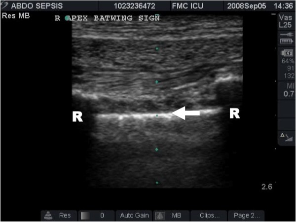
The hallmark of lung ultrasound illustrating the normal lung. The pleural line (arrow) is seen below the rib shadows (R) on either side. In real time ultrasound, lung sliding - the visual equivalent of breath sounds, can be seen as motion at the pleural line.
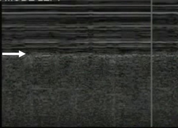
Time-motion (M-) mode ultrasonography illustrating lung sliding by the presence of the 'seashore sign'. The 'seashore sign' is characterized by motionless parietal tissue over the pleural line (arrow) and a homogenous granular pattern below it.
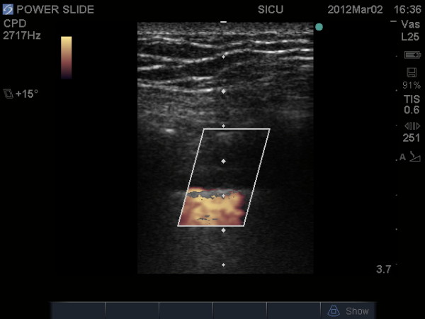
The "power slide" - Power color doppler image indicating motion at the pleural line confirming the presence of lung sliding.
Lung ultrasound image illustrating comet tail artifacts (arrow) caused by thickening of the interlobular septa.
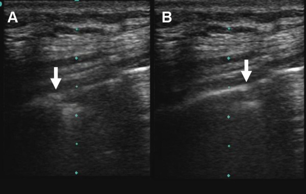
Normal pleura can be seen on the left side of the image (A) and the lung point (arrow) can be seen moving across the screen with respiration (B).
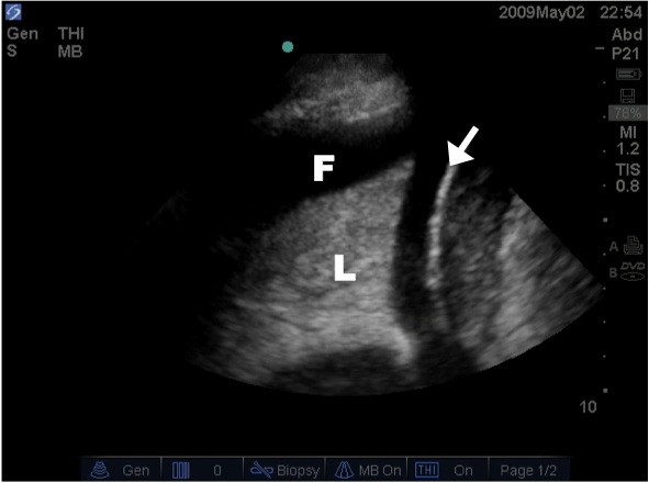
Lung ultrasound image illustrating lung consolidation, highlighted by lung (L) hepatisation ("appearing liver like").
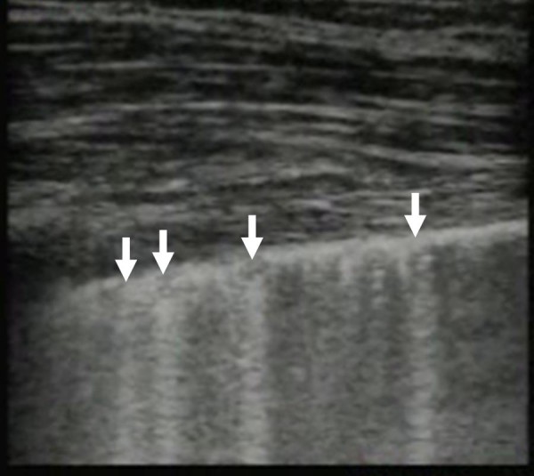
Image of the lung illustrating multiple comet tail artifacts (arrows) consistent with alveolar-interstitial syndrome, in this case caused by a pulmonary contusion.
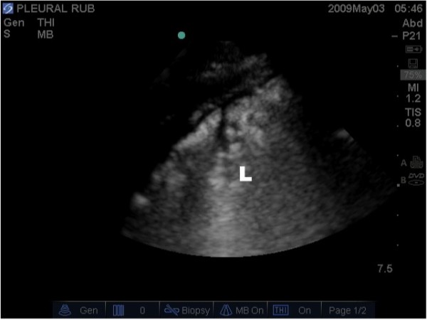
Lung ultrasound image illustrating the presence of a pleural effusion (P) around the atelectatic lung (L) above the diaphragm (arrow).
Similar articles
-
Tomorrow's stethoscope: the hand-held ultrasound device?J S C Med Assoc. 2006 Dec;102(10):345. J S C Med Assoc. 2006. PMID: 17703847 No abstract available.
-
Ten good reasons to practice ultrasound in critical care.Anaesthesiol Intensive Ther. 2014 Nov-Dec;46(5):323-35. doi: 10.5603/AIT.2014.0056. Anaesthesiol Intensive Ther. 2014. PMID: 25432552 Review.
-
Point of care ultrasonography from the emergency department to the internal medicine ward: current trends and perspectives.Intern Emerg Med. 2020 Apr;15(3):395-408. doi: 10.1007/s11739-020-02284-5. Epub 2020 Feb 7. Intern Emerg Med. 2020. PMID: 32034674 Review.
-
Pocket-size point-of-care ultrasound in rural Uganda - A unique opportunity "to see", where no imaging facilities are available.Travel Med Infect Dis. 2018 May-Jun;23:87-93. doi: 10.1016/j.tmaid.2018.01.001. Epub 2018 Jan 6. Travel Med Infect Dis. 2018. PMID: 29317333
-
Point-of-care ultrasound: Coming soon to primary care?J Fam Pract. 2018 Feb;67(2):70-80. J Fam Pract. 2018. PMID: 29400896
Cited by
-
A Comparison of Point-of-Care Ultrasonography Use in Rural Versus Urban Emergency Departments Throughout Missouri.Mo Med. 2018 Jan-Feb;115(1):56-60. Mo Med. 2018. PMID: 30228684 Free PMC article.
-
New International Guidelines and Consensus on the Use of Lung Ultrasound.J Ultrasound Med. 2023 Feb;42(2):309-344. doi: 10.1002/jum.16088. Epub 2022 Aug 22. J Ultrasound Med. 2023. PMID: 35993596 Free PMC article. Review.
-
Evaluation of musculoskeletal symptoms among physicians performing ultrasound.J Ultrason. 2017 Sep;17(70):154-159. doi: 10.15557/JoU.2017.0023. Epub 2017 Sep 29. J Ultrason. 2017. PMID: 29075519 Free PMC article.
-
Can We Place Central Venous Catheter Safely in Intensive Care Units?Indian J Crit Care Med. 2020 Jul;24(7):498-499. doi: 10.5005/jp-journals-10071-23510. Indian J Crit Care Med. 2020. PMID: 32963427 Free PMC article.
-
Integrating Ultrasound Teaching into Preclinical Problem-based Learning.J Clin Imaging Sci. 2016 Sep 20;6:38. doi: 10.4103/2156-7514.190897. eCollection 2016. J Clin Imaging Sci. 2016. PMID: 27833780 Free PMC article.
References
-
- Weinberger S, Drazen J. Harrison's Principles of Int Med. 14. New York: McGaw-Hill; 1998. Diagnostic procedures in respiratory medicine; pp. 1417–1419.
-
- Lichtenstein D, Meziere G, Biderman P, Gepner A, Barre O. The comet tail artifact: an ultrasound sign of alveolar-interstitial syndrome. Am J Respir Crit Care Med. 1997;156:1640–1646. - PubMed
-
- Volpicelli G, Caramello V, Cardinale L, Mussa A, Bar F, Frascisco MF. Detection of sonographic B-lines in patients with normal lung or radiographic alveolar consolidation. Med Sci Monit. 2008;14:CR122–CR128. - PubMed
Publication types
MeSH terms
LinkOut - more resources
Full Text Sources
Other Literature Sources
Medical
Miscellaneous
