. 2012 Mar 8:8:22.
doi: 10.1186/1746-6148-8-22.

Experimental H-type and L-type bovine spongiform encephalopathy in cattle: observation of two clinical syndromes and diagnostic challenges

Affiliations

Experimental H-type and L-type bovine spongiform encephalopathy in cattle: observation of two clinical syndromes and diagnostic challenges

Timm Konold et al. BMC Vet Res. .

Abstract

Background: The majority of atypical bovine spongiform encephalopathy (BSE) cases so far identified worldwide have been detected by active surveillance. Consequently the volume and quality of material available for detailed characterisation is very limiting. Here we report on a small transmission study of both atypical forms, H- and L-type BSE, in cattle to provide tissue for test evaluation and research, and to generate clinical, molecular and pathological data in a standardised way to enable more robust comparison of the two variants with particular reference to those aspects most relevant to case ascertainment and confirmatory diagnosis within existing regulated surveillance programmes.

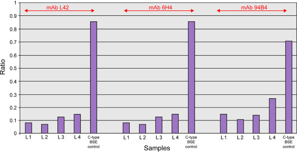
Results: Two groups of four cattle, intracerebrally inoculated with L-type or H-type BSE, all presented with a nervous disease form with some similarities to classical BSE, which progressed to a more dull form in one animal from each group. Difficulty rising was a consistent feature of both disease forms and not seen in two BSE-free, non-inoculated cattle that served as controls. The pathology and molecular characteristics were distinct from classical BSE, and broadly consistent with published data, but with some variation in the pathological characteristics. Both atypical BSE types were readily detectable as BSE by current confirmatory methods using the medulla brain region at the obex, but making a clear diagnostic distinction between the forms was not consistently straightforward in this brain region. Cerebellum proved a more reliable sample for discrimination when using immunohistochemistry.

Conclusions: The prominent feature of difficulty rising in atypical BSE cases may explain the detection of naturally occurring cases in emergency slaughter cattle and fallen stock. Current confirmatory diagnostic methods are effective for the detection of such atypical cases, but consistently and correctly identifying the variant forms may require modifications to the sampling regimes and methods that are currently in use.


Figures

Figure 1
Figure 1
Selected clinical signs observed over time in L- and H-type BSE cases compared to controls. Signs are derived from observations and examinations. The horizontal axis represents the time points when neurological examinations were carried out; a symbol at a particular time point indicates that the sign was observed between this and the previous time point. The orange dotted lines indicate the time of estimated clinical onset, the light brown interrupted lines the time of death. Head shy/restless in crush: e.g. head tossing or backing off when faced in crush. Over-reactive to tactile stimuli: e.g. head tossing in response to touching of the head. Abnormal tests of over-reactivity: startle at flash, clipboard test or hand clap, kicking on stick test. Abnormal gait: e.g. dysmetria.
Figure 2
Figure 2
Vacuolation and PrPd immunolabelling in the solitary tract nucleus of classical and atypical BSE cases. a-c) Haematoxylin and eosin (H&E) stained sections. d-f) Sections immunolabelled with monoclonal antibody (mAb) R145. Comparison of a C-type BSE case (intracerebrally inoculated case 163/95; a and d) with an L-type BSE case (L3; b and e) and an H-type BSE case (H1; c and f). In all three cases vacuolation is present, and immunolabelling is extensive and presents with particulate neuropil labelling as the predominant form.
Figure 3
Figure 3
PrPd immunolabelling in cerebellum and frontal cortex of classical and atypical BSE cases. Immunolabelling with mAb R145. a-c) Cerebellum of C-type BSE (intracerebrally inoculated case 163/95; a), L-type BSE (L1; b) and H-type BSE (H3; c). e-f) Frontal cortex of C-type BSE (163/95; d), L-type BSE (L3; e), and H-type BSE (H3; f). Of particular interest in the cerebellum is the diffuse and even involvement of both the molecular and granular layers in L-type BSE, while in the cerebellum of the H-type BSE case the most pronounced labelling is in the white matter. In the cortex small aggregated deposits, mostly in the white matter, predominate in the L-type BSE case, while the H-type BSE case resembles the classical BSE case with stellate forms throughout the cortical grey matter.
Figure 4
Figure 4
Differences in PrPd immunolabelling between L-type and H-type BSE. Immunolabelling with mAb 145, which can be used to distinguish H- and L-type forms of disease at the level of the obex. a) Labelling of the oligodendroglia in the pyramidal tract in an H-type BSE case (H3). b) Aggregated forms within the reticular formation of an L-type BSE case (L3).
Figure 5
Figure 5
Western immunoblot of medulla samples from the four H-type BSE recipients using three different antibodies. A: mAb SHA31, B: mAb SAF84, C: mAb P4. Lane Key: M Molecular mass marker. 1 Classical scrapie control 1:2 dilution. 2 C-type BSE control 1:40 dilution. 3 Blank lane. 4 H1. 5 H2. 6 H3. 7 H4.
Figure 6
Figure 6
Western immunoblot of medulla samples from the L-type BSE donor animal and the four recipients using four different antibodies. A: mAb SHA31, B: mAb 6H4, C: mAb F99, D: mAb P4. Samples were loaded neat and at dilutions indicated in the key. Lane Key: M Molecular mass marker. 1 Donor neat. 2 Donor 1:20. 3 Donor 1:40. 4 L1 neat. 5 L1 1:10. 6 L2 neat. 7 L2 1:10. 8 L3 neat. 9 L3 1:20. 10 L4 neat. 11 L4 1:10. 12 C-type BSE control neat. 13 C-type BSE control 1:20. 14 C-type BSE control 1:40. 15 Standard bovine C-type BSE control 1:40. 16 Standard ovine scrapie control 1:2.
Figure 7
Figure 7
Glycoform profiles for all four L-type BSE recipients compared to C-type BSE and scrapie using mAb SHA31. The glycoform profiles in this scattergram are displayed as the percentage signal of the diglycosylated protein band plotted against that of the monoglycosylated band. The percentage of diglycosylated PrPres is reduced in L-type BSE cases compared to C-type BSE or classical scrapie.
Figure 8
Figure 8
Bar chart of proteinase K susceptibility signal ratio in L-type recipients compared to C-type BSE using three different antibodies. The PK susceptibility signal ratio is < 0.6 for all four L-type BSE recipients and > 0.7 for the C-type BSE control sample.

References

    1. Wells GA, Scott AC, Johnson CT, Gunning RF, Hancock RD, Jeffrey M, Dawson M, Bradley R. A novel progressive spongiform encephalopathy in cattle. Vet Rec. 1987;121:419–420. doi: 10.1136/vr.121.18.419. - DOI - PubMed
    1. Wilesmith JW, Wells GA, Cranwell MP, Ryan JB. Bovine spongiform encephalopathy: epidemiological studies. Vet Rec. 1988;123:638–644. - PubMed
    1. Simmons MM, Harris P, Jeffrey M, Meek SC, Blamire IWH, Wells GAH. BSE in Great Britain: Consistency of the neurohistopathological findings in two random annual samples of clinically suspect cases. Vet Rec. 1996;138:175–177. doi: 10.1136/vr.138.8.175. - DOI - PubMed
    1. Hope J, Reekie LJD, Hunter N, Multhaup G, Beyreuther K, White H, Scott AC, Stack MJ, Dawson M, Wells GAH. Fibrils from brains of cows with new cattle disease contain scrapie-associated protein. Nature. 1988;336:390–392. doi: 10.1038/336390a0. - DOI - PubMed
    1. Stack MJ, Moore SJ, Davis A, Webb PR, Bradshaw JM, Lee YH, Chaplin M, Focosi-Snyman R, Thurston L, Spencer YI, Hawkins SAC, Arnold ME, Simmons MM, Wells GAH. Bovine spongiform encephalopathy: investigation of phenotypic variation among passive surveillance cases. J Comp Pathol. 2011;144:277–288. doi: 10.1016/j.jcpa.2010.10.007. - DOI - PubMed

Publication types

MeSH terms

LinkOut - more resources