

Long-term neuropsychological, neuroanatomical, and life outcome in hippocampal amnesia
- PMID: 22401298
- PMCID: PMC3390923
- DOI: 10.1080/13854046.2012.655781
Long-term neuropsychological, neuroanatomical, and life outcome in hippocampal amnesia
Abstract
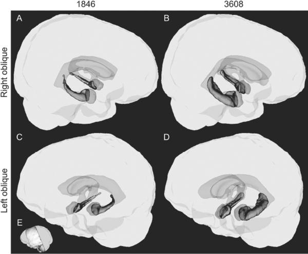
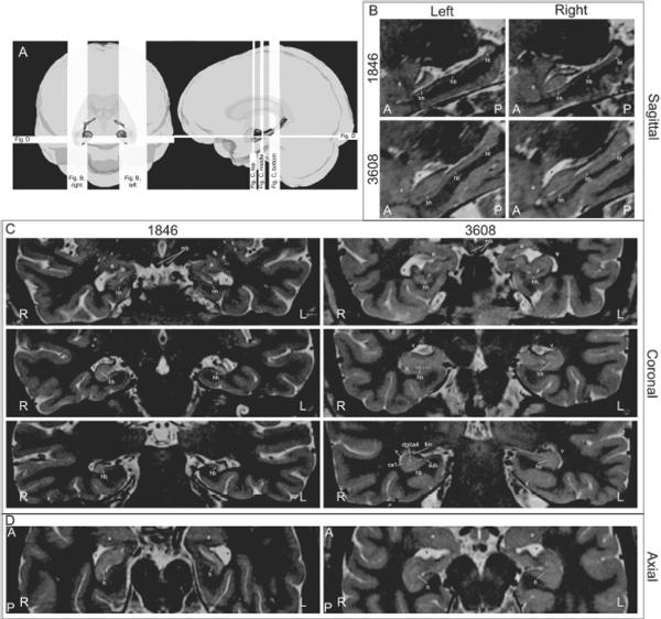
Focal bilateral hippocampal damage typically causes severe and selective amnesia for new declarative information (facts and events), a cognitive deficit that greatly impacts the ability to live a normal, fully independent life. We describe the case of 1846, a 48-year-old woman with profound hippocampal amnesia following status epilepticus and an associated anoxic episode at age 30. Patient 1846 has undergone extensive neuropsychological testing on many occasions over the 18 years since her injury, and we present data indicating that her memory impairment has remained severe and stable during that time. New, high-resolution, structural MRI studies of 1846's brain reveal substantial bilateral hippocampal atrophy resembling that of other well-known amnesic patients. In spite of severe amnesia 1846 lives a full and mostly independent adult life, facilitated by an extensive social support network of family and friends. Her case provides an example of a rare and unlikely positive outcome in the face of severe memory problems.
Figures
References
-
- Aggleton JP, Brown MW. Episodic memory, amnesia, and the hippocampal–anterior thalamic axis. Behavioral and Brain Sciences. 1999;22(3):425–489. doi:10.1017/S0140525X99002034. - PubMed
-
- Albert MS, Butters N, Brandt J. Patterns of remote memory in amnesic and demented patients. Archives of Neurology. 1981;38(8):495–500. - PubMed
-
- Albert MS, Butters N, Levin J. Temporal gradients in the retrograde amnesia of patients with alcoholic korsakoff's disease. Archives of Neurology. 1979;36(4):211–216. - PubMed
-
- Allen JS, Bruss J, Brown CK, Damasio H. Normal neuroanatomical variation due to age: The major lobes and a parcellation of the temporal region. Neurobiology of Aging. 2005;26(9):1245–60. discussion 1279-82. doi:10.1016/j.neurobiolaging.2005.05.023. - PubMed
-
- Allen JS, Tranel D, Bruss J, Damasio H. Correlations between regional brain volumes and memory performance in anoxia. Journal of Clinical and Experimental Neuropsychology. 2006;28(4):457–476. doi:10.1080/13803390590949287. - PubMed
Publication types
MeSH terms
Grants and funding
LinkOut - more resources
Full Text Sources
Medical