. 2012 Mar 8:13:4.
doi: 10.1186/1471-2121-13-4.

Rab11-FIP3 is a cell cycle-regulated phosphoprotein

Affiliations

Rab11-FIP3 is a cell cycle-regulated phosphoprotein

Louise L Collins et al. BMC Cell Biol. .

Abstract

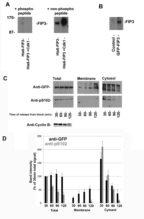
Background: Rab11 and its effector molecule, Rab11-FIP3 (FIP3), associate with recycling endosomes and traffic into the furrow and midbody of cells during cytokinesis. FIP3 also controls recycling endosome distribution during interphase. Here, we examine whether phosphorylation of FIP3 is involved in these activities.

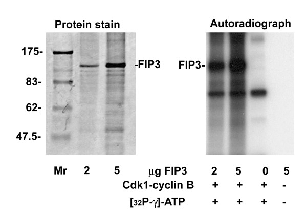
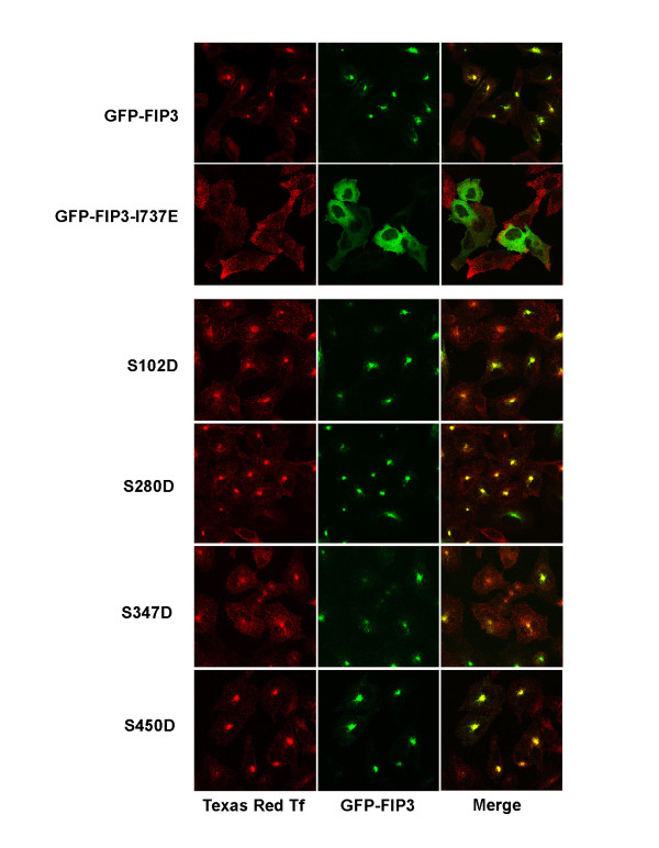
Results: We identify four sites of phosphorylation of FIP3 in vivo, S-102, S-280, S-347 and S-450 and identify S-102 as a target for Cdk1-cyclin B in vitro. Of these, we show that S-102 is phosphorylated in metaphase and is dephosphorylated as cells enter telophase. Over-expression of FIP3-S102D increased the frequency of binucleate cells consistent with a role for this phospho-acceptor site in cytokinesis. Mutation of S-280, S-347 or S-450 or other previously identified phospho-acceptor sites (S-488, S-538, S-647 and S-648) was without effect on binucleate cell formation and did not modulate the distribution of FIP3 during the cell cycle. In an attempt to identify a functional role for FIP3 phosphorylation, we report that the change in FIP3 distribution from cytosolic to membrane-associated observed during progression from anaphase to telophase is accompanied by a concomitant dephosphorylation of FIP3. However, the phospho-acceptor sites identified here did not control this change in distribution.

Conclusions: Our data thus identify FIP3 as a cell cycle regulated phosphoprotein and suggest dephosphorylation of FIP3 accompanies its translocation from the cytosol to membranes during telophase. S102 is dephosphorylated during telophase; mutation of S102 exerts a modest effect on cytokinesis. Finally, we show that de/phosphorylation of the phospho-acceptor sites identified here (S-102, S-280, S-347 and S-450) is not required for the spatial control of recycling endosome distribution or function.


Figures

Figure 1
Figure 1
FIP3 translocates from cytosol to membranes during the cell cycle Panels A and B. Synchronized HeLa cells were harvested at different time points after release from the nocodazole block as shown. Cells were fractionated into cytosolic and membrane fractions and levels of FIP3 analyzed by immunoblotting, with 60 μg of each fraction loaded in each lane. Data shown in panel A are the means and standard deviations from three independent experiments. In panel B, "prophase/metaphase" represents samples harvested at 0 min after release from nocodazole block, while "telophase" represents samples harvested 70 min after release from nocodazole block. Transferrin Receptor (TfR) was used as a marker for membranes, and RCP/FIP1 as an unrelated FIP for comparative purposes.
Figure 2
Figure 2
Identification of putative FIP3 phosphorylation sites Panel A. Synchronized HeLa cells from metaphase were lysed and FIP3 immuno-precipitated (from untreated or phosphatase-treated lysates) using anti-FIP3 antibodies. Immunoprecipitates were then separated by 2D SDS-Page and immunoblotted with anti-FIP3 antibodies. Asterisks mark different phospho-states of FIP3. A representative blot is shown for each condition. Panel B. Synchronized HeLa cells in metaphase were lysed in the presence of phosphatase inhibitors. FIP3 was then precipitated and putative phosphorylation site identified by mass spectrometry. Shown are all the peptides identified for each putative phosphorylation site. Panel C. A schematic of FIP3 showing the location of key residues. Phospho-acceptor sites identified here are shown in blue, those in black at the sites mutated to A in the HA-FIP3-4A mutant (see text). The Rab11-binding region (RBD) and Arf6 interaction region (ABD) are shown, together with the approximate position of the EF-hand-like domains and the coiled-coil region.
Figure 3
Figure 3
FIP3 is phosphorylated on S102 in a cell cycle-dependent manner. Panel A 200 ng of His6-tagged recombinant FIP3, or 200 ng His6-tagged recombinant FIP3 incubated with Cdk1-cyclin B to phosphorylate the protein on S102 (see below) were separated by SDS-PAGE, transferred to nitrocellulose and analysed by immunoblotting using 0.2 μg/ml affinity-purified anti-pS102 antibodies, incubated in the presence of either the phosphorylated or non-phosphorylated peptide (2.5 μM) as indicated. The position of molecular weight markers (kDa) is shown. Data from a typical experiment are presented, repeated four times with similar results. Panel B HeLa cells expressing GFP-FIP3 (or control cells) were lysed as described in Methods. Samples of lysate were separated by SDS-PAGE, transferred to nitrocellulose and probed with anti-pS102 antibodies. GFP-FIP3 is readily detected (endogenous FIP3 is expressed at too low levels to be readily detected by this antibody). Panel C HeLa cells stably expressing GFP-FIP3 were synchronised using a thymidine and nocodazole block as described in Methods. Cells were harvested 30, 60, 90 or 120 min after release from the block, corresponding to metaphase (~30 min) through late telophase (120 min)[11], fractionated into membrane and cytosolic fractions, and these fractions together with the initial homogenate (total) were immunoblotted with anti-GFP (to reveal total GFP-FIP3 levels in each fraction), anti-pS102 and anti-cyclin B as shown. Data from a typical experiment is presented, repeated three times with quantitatively similar results, which are quantified in Panel D (the signals at each point and in each fraction are expressed as a% of that in the 30 minute total cell lysate level. The mean of triplicate experiments (± S.D.) is shown).
Figure 4
Figure 4
Cdk1-cyclin B phosphorylates FIP3 on S102. Recombinant FIP3 was expressed as a His6-fusion protein in Sf9 cells and purified as described in Methods. The left panel shows a protein stain of 2 and 5 μg of purified FIP3 with the position of molecular weight markers indicated (kDa). 0, 2 and 5 μg of purified FIP3 were incubated with 50 ng of recombinant Cdk1-cyclin B complex in the presence of ATP-γ-32P for 30 min as described (also shown is a control in which 5 μg of FIP3 was incubated without kinase or ATP). Samples were then boiled in Laemmli buffer, separated on SDS-PAGE and examined by autoradiography. Shown is data from a typical experiment, repeated 4 times with similar results.
Figure 5
Figure 5
S102D expression perturbs cytokinesis. Panel A HeLa cells on glass coverslips were transiently transfected with GFP-FIP3, or the indicated mutants as described. After 48 h, cells were fixed and microtubules (anti-tubulin) and DNA (DAPI) stained as described. The fraction of cells expressing each FIP3 species that were binucleate was then determined and is plotted graphically. The data shown are from a representative experiment: in this case over 200 GFP-positive cells were counted per condition. *indicates a significant increase over control (GFP-FIP3) cells p = 0.02, and **p < 0.05. Similar results were obtained in two other experiments of this type, but the fraction of binucleate cells varied between experiments (e.g. the% of binucleate cells expressing GFP-FIP3-I737E varied between 16 and 33%). Panel B shows an immunoblot from the same experiment in which equal amounts of lysate were immunoblotted with anti-GFP to show that similar levels of each mutant were expressed. Anti-GAPDH is used as a loading control. Panel C shows the cytosol/membrane ratio of the indicated mutants in cells synchronised in metaphase. Data from a representative experiment is shown. Panel D HeLa cells on glass coverslips were transiently transfected with GFP-FIP3 (pseudo-coloured green), or the indicated mutants as described. After 24 h, cells were fixed and immunostained with anti-tubulin (pseudo-coloured red) and the distribution of cells in telophase examined. Shown are cells in early or late telophase. Data from a representative experiment, repeated 5 times are shown. Note that the categorisation of these images into 'early' or 'late' telophase is dictated by the appearance of FIP3 (see [11] for details) which is highly dynamic in telophase, moving from a characteristic perientrosomal localisation into the intercellular bridge region as cells move from early to late telophase. Panel E HeLa cells expressing HA-FIP3 or HA-FIP3-4A were analysed by immunofluoresence microscopy using monoclonal anti-HA in telophase to reveal the localisation of epitope-tagged FIP3. Shown are typical images of cells in early or late telophase, pseudo-coloured green. Data from a representative experiment, repeated 3 times are shown.
Figure 6
Figure 6
Distribution of phosphomimetic mutants of FIP3 and their effect on transferrin uptake in interphase cells. HeLa cells on glass coverslips were transiently transfected with GFP-FIP3 or the indicated mutants as described in Methods. 48 h later, cells were incubated for two hours in serum free D-MEM, transferred to media supplemented with 1 μg/ml human transferrin conjugated to Texas Red and incubated at 37°C for 30 minutes. Cells were fixed and the distribution of GFP-FIP3 (pseudo-coloured green) and Tf (pseudo-coloured red) analysed by confocal microscopy. Merged images were generated using the Zeiss confocal software. Shown are representative images for each mutant. Over 50 cells were analysed for each mutant at each condition, and each experiment was performed in triplicate.
Figure 7
Figure 7
Cytosol to membrane translocation of FIP3 occurs concomitantly with dephosphorylation. Panel A Synchronized HeLa cells at prophase/metaphase (0 min) or telophase (70 min) were lysed in the presence of phosphatase inhibitors as described in methods. The lysates were then passed over a Phospho-Protein purification column (Qiagen). The column was washed to remove non-phosphorylated material, and phosphoproteins eluted. The amount of FIP3 and RCP were then analyzed by immunoblotting equal amounts of lysate or eluate, as shown. Data from a representative experiment is shown, repeated with similar results. Panel B. Synchronized HeLa cells were harvested immediately after release from nocodazole block. Half of the cell lysates were treated with alkaline phosphatase before fractionation of the lysates into cytosolic and membrane fractions. The levels of FIP3, TfR and RCP in all fractions were then analyzed by immunoblotting. Data from a representative experiment is shown, repeated three times. Panel C. Density gradient analysis of FIP3 association with HeLa membranes. Phosphatase-treated, recombinant FIP3 was incubated with either HeLa membranes or protein-free liposomes. These membranes (or vesicles) were then floated up through an Optiprep gradient. Fractions were collected and the distribution of proteins within the gradients was assayed by immunoblotting using the antibodies shown. The FIP3 sample incubated with HeLa membranes was blotted for Rab5, as a marker for endocytic membranes. The bottom and top of the Optiprep density gradient is marked. Data from a typical experiment, repeated with two different preparations of lipid vesicles and HeLa membranes is shown.

References

    1. Albertson R, Riggs B, Sullivan W. Membrane traffic: a driving force in cytokinesis. Trends Cell Biol. 2005;15(2):92–101. doi: 10.1016/j.tcb.2004.12.008. - DOI - PubMed
    1. Barr FA, Greneberg U. Cytokinesis: Placing and making the final cut. Cell. 2007;131:847–860. doi: 10.1016/j.cell.2007.11.011. - DOI - PubMed
    1. Glotzer M. The molecular requirements for cytokinesis. Science. 2005;307:1735–1739. doi: 10.1126/science.1096896. - DOI - PubMed
    1. Prekeris R, Gould GW. Breaking up is hard to do: Membrane traffic in cytokinesis. J Cell Sci. 2008;121:1569–1576. doi: 10.1242/jcs.018770. - DOI - PMC - PubMed
    1. Schiel JA, Prekeris R. Making the final cut - mechanisms mediating the abscission step of cytokinesis. Scientific World Journal. 2010;10:1424–1434. - PMC - PubMed

Publication types

LinkOut - more resources