. 2012 Mar 9:9:5.
doi: 10.1186/1476-9255-9-5.

STAT6 expression in T cells, alveolar macrophages and bronchial biopsies of normal and asthmatic subjects

Affiliations

STAT6 expression in T cells, alveolar macrophages and bronchial biopsies of normal and asthmatic subjects

Katsuyuki Tomita et al. J Inflamm (Lond). .

Abstract

Background: Asthma is characterised by increased numbers of Th2-like cells in the airways and IgE secretion. Generation of Th2 cells requires interleukin (IL)-4 and IL-13 acting through their specific receptors and activating the transcription factor, signal transducer and activator of transcription 6 (STAT6). STAT6 knockout mice fail to produce IgE, airway hyperresponsiveness and bronchoalveolar lavage eosinophilia after allergen sensitisation, suggesting a critical role for STAT6 in allergic responses.

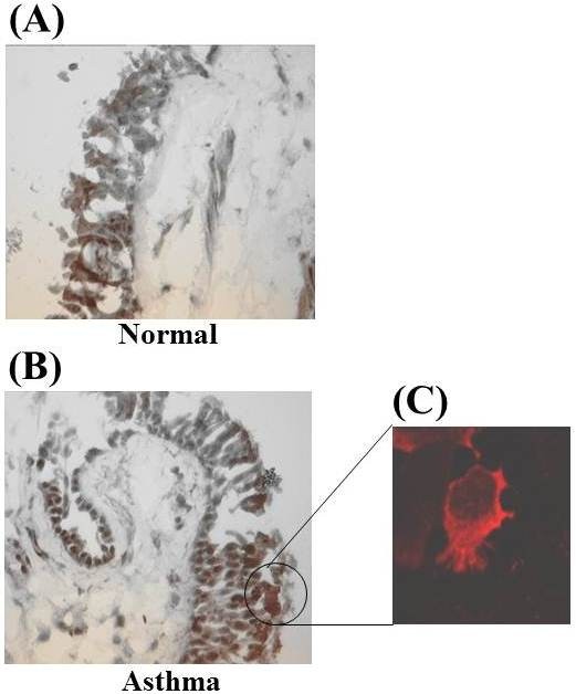
Methods: We have investigated the expression of STAT6 in peripheral blood T-lymphocytes, alveolar macrophages and bronchial biopsies from 17 normal subjects and 18 mild-moderate steroid-naïve stable asthmatic patients.

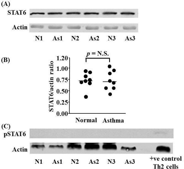
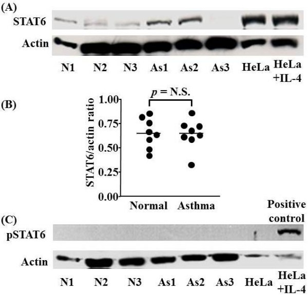
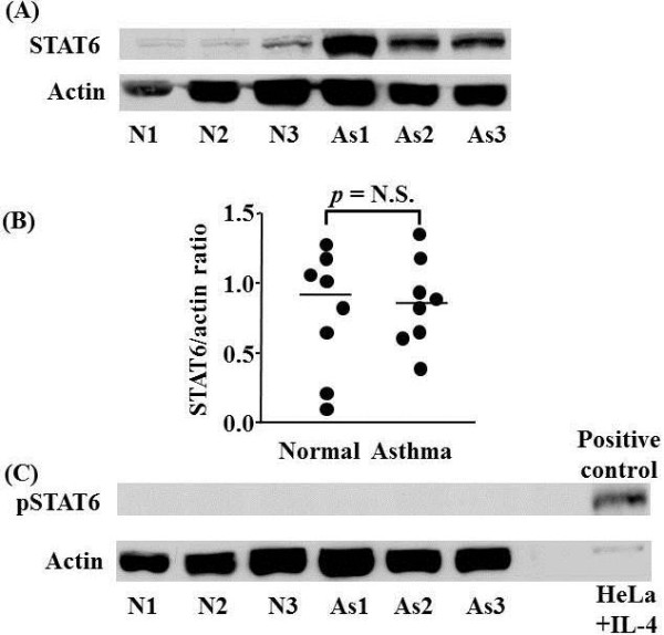
Results: STAT6 expression was variable and was detected in T-lymphocytes, macrophages and bronchial epithelial cells from all subjects with no difference between normal and stable asthmatic subjects.

Conclusions: STAT6 expression in different cells suggests that it may be important in regulating the expression of not only Th2-like cytokines in T cells of man, but may also regulate STAT-inducible genes in alveolar macrophages and airway epithelial cells.


Figures

Figure 1
Figure 1
STAT6 protein expression in peripheral blood T-lymphocytes isolated from asthmatic (As) and normal (N) subjects. Representative Western blot analyses of experiments from 3 individual subjects in each group are shown (A). Actin expression is used to control for protein loading. Graphical representation of the data in (A) is shown in (B). (C) Failure to detect phospho-STAT6 (pSTAT6) protein expression in peripheral blood T-lymphocytes isolated from As and N subjects. pSTAT6 expression in control Th2 cells confirms the ability of the antibody to detect pSTAT6. Each circle represents one subject. Horizontal bars represent median values.
Figure 2
Figure 2
Representative immunocytochemical staining for STAT6 in T-lymphocytes (A), CCR5- (B) and CCR5+ cells (C). STAT6 is localised predominantly to the cytoplasm. However, in a small subset of T cells nuclear localisation of STAT6 can be detected. STAT6 is localised predominantly to the nucleus of CCR5- but not CCR5+ cells.
Figure 3
Figure 3
Representative Western blot analysis of STAT6 and phospho-STAT6 protein expression in alveolar macrophages of normal (N) and asthmatic (As) subjects. Representative Western blot analyses of experiments from 3 individual subjects in each group are shown (A). Actin expression is used to control for protein loading. Graphical representation of the data in (A) is shown in (B). (C) Failure to detect phospho-STAT6 (pSTAT6) protein expression in alveolar macrophages isolated from As and N subjects. pSTAT6 expression in control IL-4 stimulated HeLa cells confirms the ability of the antibody to detect pSTAT6. Each circle represents one subject. Horizontal bars represent median values.
Figure 4
Figure 4
Western blot analysis of STAT6 and phospho-STAT6 expression in bronchial mucosal biopsies of normal (N) and asthmatic (As) subjects. Representative Western blot analyses of experiments from 3 individual subjects in each group are shown (A). Actin expression is used to control for protein loading. Graphical representation of the data in (A) is shown in (B). (C) Failure to detect phospho-STAT6 (pSTAT6) protein expression in bronchial biopsies isolated from As and N subjects. pSTAT6 expression in control IL-4 stimulated HeLa cells confirms the ability of the antibody to detect pSTAT6. Each circle represents one subject. Horizontal bars represent median values.
Figure 5
Figure 5
Immunohistochemical staining for STAT6 protein in bronchial mucosal biopsies of normal (A) and asthmatic (B) subject, and representative immunofluorescence staining for STAT6 in bronchial epithelial cells of an asthmatic subject (C).It indicates that STAT6 is localised predominantly to the cytoplasm in bronchial cells of an asthmatic subject.

Similar articles

Cited by

References

    1. Barnes PJ, Chung KF, Page CP. Inflammatory mediators of asthma: an update. Pharmacol Rev. 1998;50:515–596. - PubMed
    1. Hart L, Lim S, Adcock I, Barnes PJ, Chung KF. Effects of inhaled corticosteroid therapy on expression and DNA-binding activity of nuclear factor kappaB in asthma. Am J Respir Crit Care Med. 2000;161:224–231. - PubMed
    1. Hart LA, Krishnan VL, Adcock IM, Barnes PJ, Chung KF. Activation and localization of transcription factor, nuclear factor-kappaB, in asthma. Am J Respir Crit Care Med. 1998;158:1585–1592. - PubMed
    1. Demoly P, Basset SN, Chanez P. et al.c-fos proto-oncogene expression in bronchial biopsies of asthmatics. Am J Respir Cell Mol Biol. 1992;7:128–133. - PubMed
    1. Annunziato F, Galli G, Cosmi L, Romagnani P, Manetti R, Maggi E, Romagnani S. Molecules associated with human Th1 or Th2 cells. Eur Cytokine Netw. 1998;9:12–16. - PubMed