Review
. 2012 Apr;22(4):201-10.
doi: 10.1016/j.tcb.2012.02.001. Epub 2012 Mar 6.

The ciliary transition zone: from morphology and molecules to medicine

Affiliations
Review

The ciliary transition zone: from morphology and molecules to medicine

Peter G Czarnecki et al. Trends Cell Biol. 2012 Apr.

Abstract

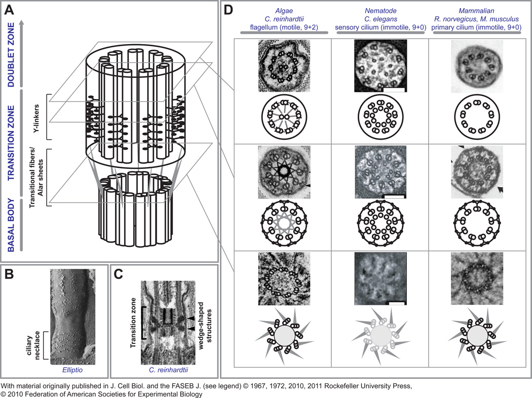
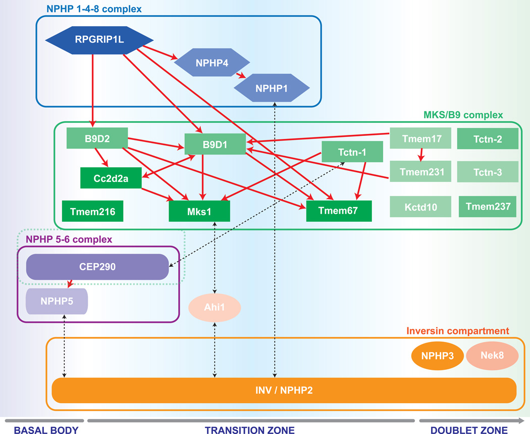
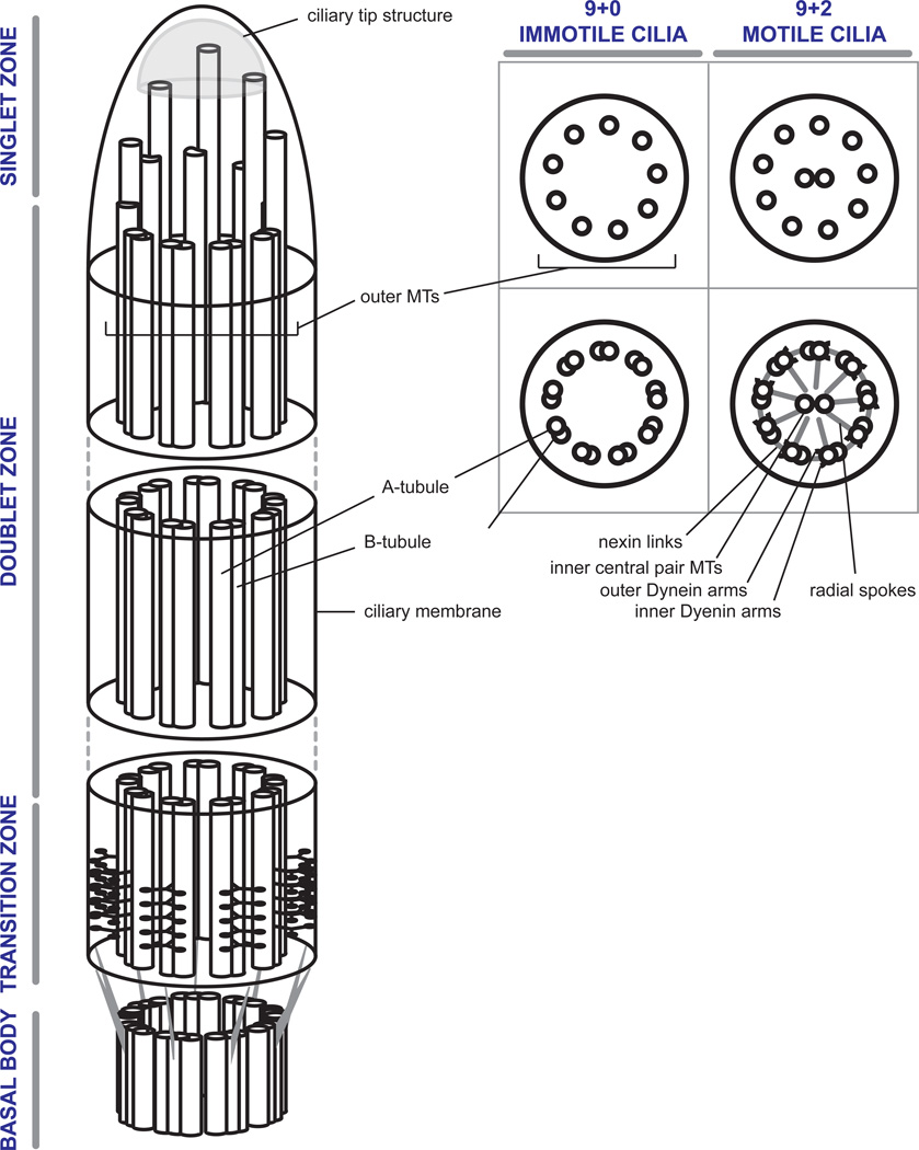
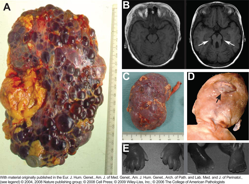
Researchers from various disciplines, including cell and developmental biology, genetics and molecular medicine, have revealed an exceptional diversity of cellular functions that are mediated by cilia-dependent mechanisms. Recent studies have directed our attention to proteins that localize to the ciliary transition zone (TZ), a small evolutionarily conserved subcompartment that is situated between the basal body (BB) and the more distal ciliary axoneme. These reports shed light on the roles of TZ proteins in ciliogenesis, ciliary protein homeostasis and specification of ciliary signaling, and pave the way for understanding their contribution to human ciliopathies. In this review, we describe the interplay of multimeric protein complexes at the TZ, integrating morphological, genetic and proteomic data towards an account of TZ function in ciliary physiology.


Figures

Figure 1
Figure 1. The Ciliary Transition Zone
A – A schematic representation of the ciliary transition zone shows its location between the basal body compartment and the doublet zone of the axoneme. At the distal end of the basal body, terminal triplet MT architecture and transitional fibers are seen. The A–B tubules of the triplet MTs are continuous with the doublets (not shown here for clarity) within the transition zone proper where characteristic Y-linkers span from the doublets to the surrounding membrane. It is thought that these interactions produce characteristic ciliary membrane imprints referred to as the ciliary necklace (B, reproduced with permission from [5]). In the transition zone of Chlamydomonas flagella, there is an additional central density with a barrel-shaped body within the space defined by the MT doublets and a wedge shaped density projecting towards the membrane from the outer MTs (C, reproduced with permission from [10]). D - Although remarkably conserved, subtle structural differences exist between model organisms. At the start of the transition zone, transitional fibers project from the B-tubule to the ciliary membrane at its junction with the apical cell membrane. The appearance of transitional fibers indicate the distal end of the basal body, since at that point, C-tubules terminate and the transition zone and axoneme consist only of outer MT doublets. Transitional fibers, also called alar sheets, form a propeller-like assembly leaving spaces of about 60 nm between neighboring sheets. The BB structures (grayed MT triplets) are only poorly discerned in Caenorhabditis sensory cilia, but transitional fibers can be observed. Within the transition zone, all cilia have the characteristic Y-linkers connecting the outer doublet MTs to the ciliary membrane. In addition, Chlamydomonas has a stellate fiber array within the outer doublet array connected to all A-tubules that appears near the basal plate. Within the axoneme of motile cilia, such as in Chlamydomonas, an arrangement of radial spoke proteins connect the inner central pair of MTs to the outer doublets, and nexin fibers and dynein arms connect the outer doublets to each other. The sensory cilia of Caenorhabditis and the primary cilium of mammalian cells both lack the inner central pair and the basal plate from which they originate. In addition, they lack radial spokes, nexin fibers and dynein arms (reproduced with permission from [10, 11, 30, 41, 42]). Scale bars, 100 nm.
Figure 2
Figure 2. Organizational chart of transition zone functional modules
Rpgrip1L/NPHP8 localizes independently and influences proper TZ localization of the NPHP1-4 and MKS/B9 complexes. The localization may be direct or through a cascade of consecutive localization and complex organization events. Within the MKS/B9 module, Mks1, Tmem216 and Tmem67, which are consistently associated with severe pathologies in humans, appear at the bottom of the localization hierarchy within the module. Cep290 was considered the core of its own NPHP5-6 network in one study [15], but part of an MKS-like complex in another [11]. The inversin compartment is highly dynamic and spans a long stretch of ciliary axoneme from the basal body into the distal cilium proper. Through its localization and its interactions with members of all protein modules, it may serve as a bridge and contribute to multiple distinct signaling processes. Proteins within one module are highlighted by the same shape and color. Brighter color indicates more frequent association with severe disease. Higher position within one group and red arrows indicate intra-complex localization hierarchy. Black dotted arrows indicate physical interactions across modules. Note that not all depicted proteins are exclusively located in the TZ, but may also be found at the basal body (Rpgrip1L, B9D1, Cc2d2a, Tmem216, Cep290, NPHP5, inversin), or in the proximal doublet zone of the cilium proper (inversin, Nphp3, Nek8, Tmem67, Tctn-2, Tctn-3).
Box 1
Box 1
Box 2
Box 2

References

    1. Carvalho-Santos Z, et al. Evolution: Tracing the origins of centrioles, cilia, and flagella. The Journal of Cell Biology. 2011;194:165–175. - PMC - PubMed
    1. Fisch C, Dupuis-Williams P. Ultrastructure of cilia and flagella - back to the future! Biology of the cell / under the auspices of the European Cell Biology Organization. 2011;103:249–270. - PubMed
    1. Anderson RG. The three-dimensional structure of the basal body from the rhesus monkey oviduct. The Journal of Cell Biology. 1972;54:246–265. - PMC - PubMed
    1. Gibbons IR, Grimstone AV. On flagellar structure in certain flagellates. The Journal of biophysical and biochemical cytology. 1960;7:697–716. - PMC - PubMed
    1. Gilula NB, Satir P. The ciliary necklace. A ciliary membrane specialization. The Journal of Cell Biology. 1972;53:494–509. - PMC - PubMed

Publication types