. 2012 Apr 15;205(8):1203-13.
doi: 10.1093/infdis/jis102. Epub 2012 Mar 7.

The Mycobacterium tuberculosis stress response factor SigH is required for bacterial burden as well as immunopathology in primate lungs

Affiliations

The Mycobacterium tuberculosis stress response factor SigH is required for bacterial burden as well as immunopathology in primate lungs

Smriti Mehra et al. J Infect Dis. .

Abstract

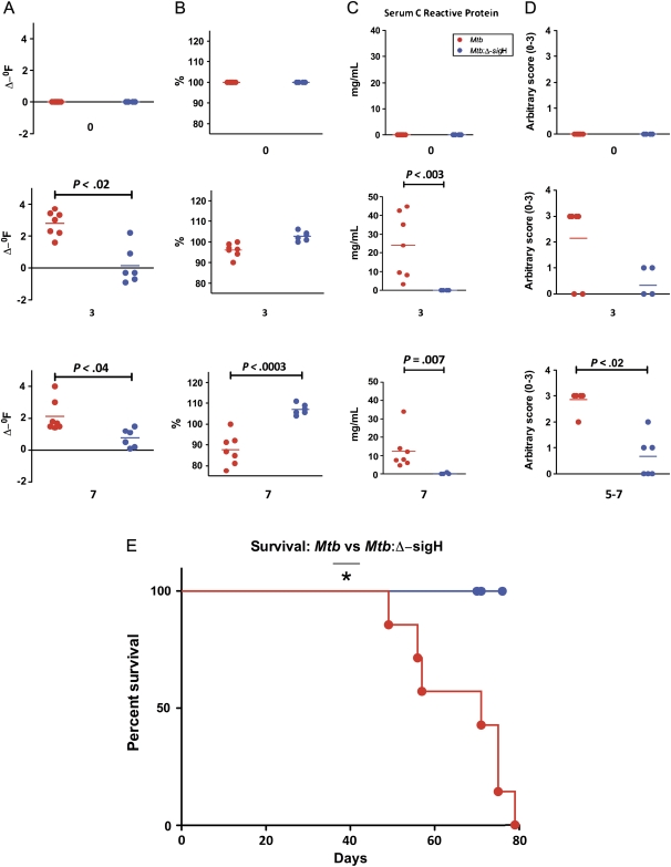
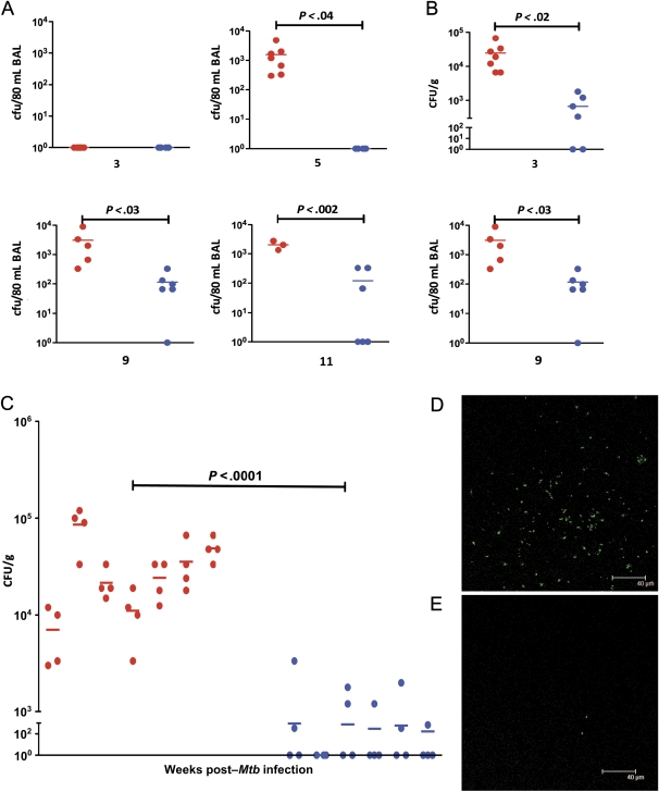
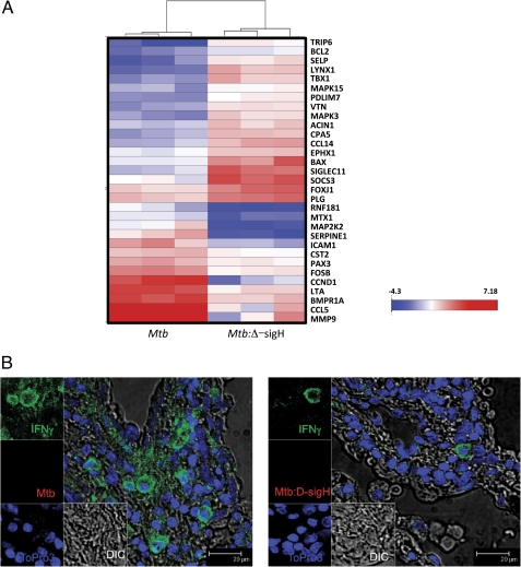
Background: Sigma H (sigH) is a major Mycobacterium tuberculosis (Mtb) stress response factor. It is induced in response to heat, oxidative stress, cell wall damage, and hypoxia. Infection of macrophages with the Δ-sigH mutant generates more potent innate immune response than does infection with Mtb. The mutant is attenuated for pathology in mice.

Methods: We used a nonhuman primate (NHP) model of acute tuberculosis, to better understand the phenotype of the Δ-sigH mutant in vivo. NHPs were infected with high doses of Mtb or the mutant, and the progression of tuberculosis was analyzed in both groups using clinical, pathological, microbiological, and immunological parameters.

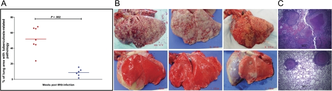
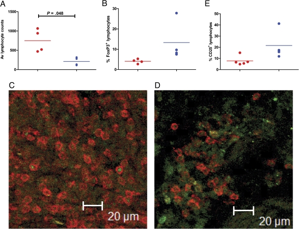
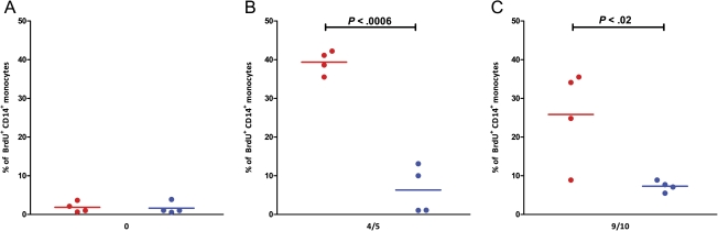
Results: Animals exposed to Mtb rapidly progressed to acute pulmonary tuberculosis as indicated by worsening clinical correlates, high lung bacterial burden, and granulomatous immunopathology. All the animals rapidly succumbed to tuberculosis. On the other hand, the NHPs exposed to the Mtb:Δ-sigH mutant did not exhibit acute tuberculosis, instead showing significantly blunted disease. These NHPs survived the entire duration of the study.

Conclusions: The Mtb:Δ-sigH mutant is completely attenuated for bacterial burden as well as immunopathology in NHPs. SigH and its regulon are required for complete virulence in primates. Further studies are needed to identify the molecular mechanism of this attenuation.


Figures

Figure 1.
Figure 1.
Clinical data from nonhuman primates (NHPs) infected with Mycobacterium tuberculosis (Mtb) as well as the Mtb:Δ-sigH mutant. A, Changes in body temperature, expressed in Δ-°F. B, Changes in body weight, expressed as percentage of total weight at the time of Mtb infection. C, Changes in serum C-reactive protein (CRP) levels. D, Changes in arbitrary chest radiographic (CXR) scores. E, Survival proportions. Data are shown for week 0 (preinfection) as well as weeks 3 and 7 postinfection for A, B, C, and D. The x-axis values represent week postinfection. Red circles denote NHPs infected with Mtb, whereas blue circles denote NHPs infected with the Mtb:Δ-sigH mutant. Significant differences are shown wherever detected.
Figure 2.
Figure 2.
Bacterial burden in the 2 groups of nonhuman primates (NHPs). Temporal Mycobacterium tuberculosis (Mtb) colony-forming units (CFUs) are shown per 80 mL bronchoalveolar lavage (BAL) samples obtained from both groups of NHPs at weeks 3, 5, and 9 postinfection. A, Temporal Mtb CFUs are shown per gram of bronchial lymph node tissue obtained from both groups of NHPs at weeks 3 and 7 postinfection (B). Mtb colony-forming units (CFUs) are also shown per gram of lung tissue obtained at necropsy from both groups of NHPs (C). Each lung was randomly sectioned at necropsy and 10 sections from each of the 2 lungs were pooled into 2 groups (right lung 1, right lung 2; left lung 1, left lung 2). Results are shown for all 4 of these lung samples for each of the 13 NHPs. The x-axis values represent the week postinfection. Red circles denote NHPs infected with Mtb, and blue circles denote NHPs infected with the Mtb:Δ-sigH mutant. Confocal microscopy shows the extent of bacterial presence in the lungs of a representative NHP infected with Mtb (D) relative to a representative NHP infected with Mtb:Δ-sigH (E).
Figure 3.
Figure 3.
Postnecropsy pathology data from the 2 groups of nonhuman primates (NHPs). The percentage of lung area with tuberculosis-related pathology was estimated in both groups of NHPs (A) using methods described elsewhere [23, 25]. The x-axis values represent week postinfection. Red circles denote NHPs infected with Mycobacterium tuberculosis (Mtb), whereas blue circles denote NHPs infected with the Mtb:Δ-sigH mutant. Lines correspond to mean values. The differences between 2 groups were statistically significant. Gross pathology is shown for 3 representative NHPs each, infected with Mtb (top panel) and Mtb:Δ-sigH (bottom panel) (B). Histopathologic analysis of hematoxylin and eosin–stained lung samples from 1 representative NHP each, infected with Mtb (top panel) and Mtb:Δ-sigH (bottom panel) (C).
Figure 4.
Figure 4.
Immune response analysis using microarrays and confocal microscopy. Triplicate RNA samples derived from tuberculosis lesions of 3 nonhuman primates (NHPs) of each group were profiled on Agilent Rhesus Macaque 4 × 44 microarrays. The derived expression changes were sorted based on significance. Data are shown as heat-map clusters for genes with highly significantly differential expression in NHPs infected with Mycobacterium tuberculosis (Mtb) or the Mtb:Δ-sigH relative to the other group (A). The color schematics for the heat map are as follows: blue, lower expression relative to normal lung; white, comparable expression relative to normal lung; red, higher expression relative to normal lung. The intensity of red and blue color corresponds to the extent of induction or repression and is shown as a color bar (A). Multilabel confocal microscopy shows differential expression of the foremost anti-Mtb Th1 type proinflammatory cytokine interferon γ (IFN-γ) in the lung lesion of an NHP infected with Mtb (B) and Mtb:Δ-sigH (C). Eukaryotic cells were detected using a nuclear stain (TO-PRO3) (blue signal), and Mtb was detected using an Mtb-specific antibody (red signal), both as described elsewhere [–26]. IFN-γ was detected using a specific antibody (green signal) as described in the “Materials and Methods” section.
Figure 5.
Figure 5.
Identification of regulatory T cells (Tregs) in the tubercle lesions of the 2 groups of nonhuman primates (NHPs). Immunohistochemisty (AC) and immunofluorescence were performed to identify total CD3+ T cells (A, D), CD3+ FoxP3+Tregs (B, E), and CD3+CD25+ activated Tregs (E). For immunohistochemical determination of total numbers, 6 fields per slide were counted for 4 NHPs in each group at ×20 magnification using a Leica DMLB scope and a SPOT Insight Color 3.2.0 camera. Images were taken with SPOT 3.4.5 software and counted using Image Pro-Plus 4.5.0.19.
Figure 6.
Figure 6.
In vivo bromodeoxyuridine (BrdU) labeling and flow cytometry. Four animals from each group were injected with BrdU (10 mg/kg body weight) as described elsewhere [28], prior to infection (A) and 4–5 weeks postinfection (B). Blood was collected 24 hours after BrdU inoculation. The percentages of BrdU-positive cells in CD14+ monocytes were analyzed by flow cytometry as described elsewhere [28].

Comment in

References

    1. Raviglione MC. The new Stop TB strategy and the Global Plan to Stop TB, 2006–2015. Bull World Health Organ. 2007;85:327. - PMC - PubMed
    1. Gandhi NR, Moll A, Sturm AW, et al. Extensively drug-resistant tuberculosis as a cause of death in patients co-infected with tuberculosis and HIV in a rural area of South Africa. Lancet. 2006;368:1575–80. - PubMed
    1. Adegbola RA, Hill PC, Secka O, et al. Surveillance of drug-resistant Mycobacterium tuberculosis in The Gambia. Int J Tuberc Lung Dis. 2003;7:390–3. - PubMed
    1. Corbett EL, Watt CJ, Walker N, et al. The growing burden of tuberculosis: global trends and interactions with the HIV epidemic. Arch Intern Med. 2003;163:1009–21. - PubMed
    1. Fine PE. Variation in protection by BCG: implications of and for heterologous immunity. Lancet. 1995;346:1339–45. - PubMed

Publication types

MeSH terms