Comparative Study
. 2012 May;32(5):1246-54.
doi: 10.1161/ATVBAHA.111.241257. Epub 2012 Mar 8.

Metalloproteinase processing of HBEGF is a proximal event in the response of human aortic endothelial cells to oxidized phospholipids

Affiliations
Comparative Study

Metalloproteinase processing of HBEGF is a proximal event in the response of human aortic endothelial cells to oxidized phospholipids

Sangderk Lee et al. Arterioscler Thromb Vasc Biol. 2012 May.

Abstract

Objective: Atherosclerosis is a chronic inflammatory disease initiated by monocyte recruitment and retention in the vessel wall. An important mediator of monocyte endothelial interaction is the chemokine interleukin (IL)-8. The oxidation products of phospholipids, including oxidized 1-palmitoyl-2-arachidonyl-sn-glycerol-3-phosphocholine (Ox-PAPC), accumulate in atherosclerotic lesions and strongly induce IL-8 in human aortic endothelial cells (HAECs). The goal of this study was to identify the proximal events leading to induction of IL-8 by Ox-PAPC in vascular endothelial cells.

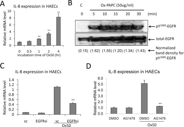
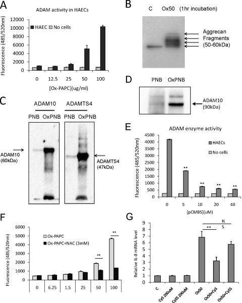
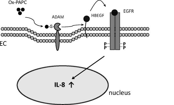
Methods and results: In a systems genetics analysis of HAECs isolated from 96 different human donors, we showed that heparin-binding EGF-like growth factor (HBEGF) transcript levels are strongly correlated to IL-8 induction by Ox-PAPC. The silencing and overexpression of HBEGF in HAECs confirmed the role of HBEGF in regulating IL-8 expression. HBEGF has been shown to be stored in an inactive form and activation is dependent on processing by a dysintegrin and metalloproteinases (ADAM) to a form that can activate the epidermal growth factor (EGF) receptor. Ox-PAPC was shown to rapidly induce HBEGF processing and EGF receptor activation in HAECs. Using siRNA we identified 3 ADAMs that regulate IL-8 induction and directly demonstrated that Ox-PAPC increases ADAM activity in the cells using a substrate cleavage assay. We provide evidence for one mechanism of Ox-PAPC activation of ADAM involving covalent binding of Ox-PAPC to cysteine on ADAM. Free thiol cysteine analogs showed inhibition of IL-8 induction by Ox-PAPC, and both a cysteine analog and a cell surface thiol blocker strongly inhibited ADAM activity induction by Ox-PAPC. Using microarray analyses, we determined that this ADAM pathway may regulate at least 30% of genes induced by Ox-PAPC in HAECs.

Conclusions: This study is the first report demonstrating a role for the ADAM-HBEGF-EGF receptor axis in Ox-PAPC induction of IL-8 in HAECs. These studies highlight a role for specific ADAMs as initiators of Ox-PAPC action and provide evidence for a role of covalent interaction of Ox-PAPC in activation of ADAMs.


Figures

Figure 1
Figure 1. Ox-PAPC induced IL-8 expression shows a high level of variation in HAECs isolated from 96 human donors
HAECs were treated as duplicate with media (C) or Ox-PAPC (50ug/ml) for 4 hrs, and the IL-8 transcript levels were determined by microarray analysis as described in the method section. The intensity of signalings before (○) and after (●) Ox-PAPC treatment are shown on the Y axis, and the cell lines are shown along the X axis. The lines have been rank-ordered on the base of basal levels. Arrows indicate representative cell lines showing strong and weak induction of IL-8.
Figure 2
Figure 2. IL-8 induction is strongly regulated by HBEGF
(A) HBEGF silencing decreased IL-8 induction by Ox-PAPC in HAECs. (B–C) Overexpression of HBEGF in HAECs (B) or the introduction of HBEGF in HEK293 cells (C) increased IL-8 expression induced by Ox-PAPC. Endogenous HBEGF level was undetectable in untransfected HEK293 cells. After 2 days following transfection of siRNA or HBEGF plasmid, cells were treated with Ox-PAPC (50ug/ml) for 4hrs and the IL-8 mRNA levels were determined by qRT-PCR. Western blotting and qRT-PCR were repeated at least three times and representative results shown. The silencing of target proteins was confirmed by qRT-PCR as shown in Suppl. Figure I. (D) Active HBEGF is increased in response to Ox-PAPC. HAECs were treated with Ox-PAPC (50ug/ml) for 0, 10, and 30min in duplicate, and the proteins in cell lysates were separated using SDS-PAGE. The formation of active HBEGF was determined by Western blotting. (E) Recombinant HBEGF significantly increased IL-8 in HAECs. HAECs were treated with HBEGF for 4 hrs and the levels of IL-8 were determined by qRT-PCR. The values were represented as mean + SD, *p<0.05 and ** p<0.01, N=3 for reactions.
Figure 3
Figure 3. EGFR mediates the induction of IL-8 by Ox-PAPC in HAECs
(A) Ox-PAPC increased IL-8 expression in HAECs in a time-dependent manner as determined by qRT-PCR. (B) Ox-PAPC activates EGFR. Cells were treated with Ox-PAPC (50ug/ml) for 0 to 30 min and the phosphorylation of Tyr1068 of EGFR was determined by Western blot using pY1068 -EGFR antibody. (C) EGFR silencing by siRNA transfection significantly reduced IL-8 induction by Ox-PAPC (50ug/ml, 4hrs). The mRNA content of IL-8 was determined by qRT-PCR. Multiple siRNAs for EGFR with different nucleotide sequences showed similar effects. The silencing of target proteins was confirmed by qRT-PCR and Western blotting as shown in Suppl. Figure II. (D) EGFR inhibitor reduced IL- expression by Ox-PAPC. Cells were pretreated with EGFR inhibitor AG1498 (10uM) for 1hr, and cotreated with Ox-PAPC (50ug/ml) for 4hrs. Vehicle (DMSO) was used as control treatment. The IL-8 mRNA levels were determined by qRT-PCR. Western blotting and qRT-PCR were repeated at least three times with reproducibility and representative results were shown. The values were represented as mean+SD, ** p<0.01, N=3 for reaction.
Figure 4
Figure 4. Specific ADAMs regulates IL-8 induction by Ox-PAPC in HAECs
(A) Metalloproteinase mediates IL-8 expression in HAECs. HAECs were pretreated with the pan-metalloproteinase inhibitor GM6001 (10uM) for 1hr and co-treated with Ox-PAPC (50ug/ml, Ox50) for 4hrs, and the mRNA levels of IL-8 were determined by qRT-PCR. (B) Metalloproteinase mediates EGFR activation by Ox-PAPC in HAECs. Metalloproteinase inhibitor TAPI-1 reduced EGFR activation by Ox-PAPC in HAECs. Cells were pretreated with TAPI-1 (10uM) for 1 hr and co-treated with Ox-PAPC (50ug/ml) for 20min, and the relative EGFR activation was determined by Western blotting using pY1068 specific -EGFR antibody. (C, D) ADAM10, ADAM19, and ADAMTS4 mediate IL-8 expression by Ox-PAPC in HAECs. Gene silencing of ADAM10, ADAM19, and ADAMTS4 showed significant reductions in IL-8 expression by Ox-PAPC in HAECs. HAECs were treated and qRT-PCR performed as in (A). The silencing of target proteins was confirmed by qRT-PCR and Western blotting as shown in Suppl. Figures IV and V. Cell treatment and qRT-PCR were repeated at least three times and representative results shown. The values were mean values + SD, ** p<0.01, N=3 for each PCR reaction.
Figure 5
Figure 5. Ox-PAPC increases ADAM activities in HAECs and covalently interacts with ADAMs
(A) Ox-PAPC dose-dependently increased ADAM activity in HAECs. HAEC were treated for 1 hour with different doses of Ox-PAPC and ADAM activity in the cells was determined using fluorogenic ADAM substrate as described in Methods section. (B) Ox-PAPC induced the digestion of endogenous ADAM-TS substrate aggrecan in HAECs. HAECs were treated with Ox-PAPC (50ug/ml) for 1hr, and the content of aggrecan fragments in the cell supernatants were determined by Western blotting using aggrecan-specific antibody. (C) Oxidized phospholipids covalently interact with recombinant ADAM10 and ADAMTS4 commercially available. Active form of ADAM10 and ADAMTS4 (1ug) were incubated with unoxidized and oxidized PAPE-N-biotin (50ug/ml) for 1 hr, and the reactions were run in SDS-PAGE in reducing condition. The presence of biotin-labeled protein was detected by probing with streptavidin-HRP. (D) Oxidized phospholipids also covalently interact with endogenous ADAM10 in HAECs. HAECs were incubated with unoxidized and oxidized PAPE-N-biotin (50ug/ml) for 1 hr, and the lysates in RIPA buffer were incubated with avidin-beads overnight for precipitation. After stringent washing with RIPA, the presence of ADAM10 biotin-labeled was detected by probing with ADAM10-specific antibody by Western blotting. (E) Cell surface free thiol is required for ADAM activation by Ox-PAPC in HAECs. HAECs were pretreated with differential doses of cell surface free thiol blocker p-chloromercuribenzene sulfonate (pCMBS) and Ox-PAPC (50ug/ml) for 1hr, and and ADAM activity as described in (A). The ranges of pCMBS used in this study did not show any obvious cell damage in HAECs. (F) The cysteine analog N-acetyl-L-cysteine (NAC) interacts with Ox-PAPC and inhibits ADAM activation in HAECs. HAECs were pretreated with different doses of NAC for 1hr in the presence of Ox-PAPC (50ug/ml), and ADAM activity as described in (A). (G) HAECs were pretreated with 200uM of cysteine or cystine for 1hr and cotreated with control media or Ox-PAPC (50ug/ml) for 4 hrs. The levels of IL-8 were determined by qRT-PCR. Western blotting and enzyme assay were repeated at least three times with reproducibility and representative results were shown. The values were represented as mean value + SD, ** p<0.01, N=3 for PCR or enzyme reactions.
Figure 6
Figure 6. Hypothetical diagram depicting the proximal steps of Ox-PAPC signaling of IL-8 induction in vascular endothelial cells
Ox-PAPC activates ADAMs on the vascular endothelial cell surface, and the activated ADAM processes HBEGF for EGFR activation, leading to IL-8 expression in the nucleus.

References

    1. Steinberg D, Witztum JL. Oxidized low-density lipoprotein and atherosclerosis. Arterioscler Thromb Vasc Biol. 2010;30:2311–2316. - PubMed
    1. Levitan I, Volkov S, Subbaiah PV. Oxidized ldl: Diversity, patterns of recognition, and pathophysiology. Antioxid Redox Signal. 2010;13:39–75. - PMC - PubMed
    1. Bochkov VN, Oskolkova OV, Birukov KG, Levonen AL, Binder CJ, Stockl J. Generation and biological activities of oxidized phospholipids. Antioxid Redox Signal. 2010;12:1009–1059. - PMC - PubMed
    1. Yla-Herttuala S, Palinski W, Rosenfeld ME, Parthasarathy S, Carew TE, Butler S, Witztum JL, Steinberg D. Evidence for the presence of oxidatively modified low density lipoprotein in atherosclerotic lesions of rabbit and man. J Clin Invest. 1989;84:1086–1095. - PMC - PubMed
    1. Berliner JA, Leitinger N, Tsimikas S. The role of oxidized phospholipids in atherosclerosis. J Lipid Res. 2009;50(Suppl):S207–212. - PMC - PubMed

Publication types

MeSH terms