

In vivo proton dosimetry using a MOSFET detector in an anthropomorphic phantom with tissue inhomogeneity
- PMID: 22402385
- PMCID: PMC5716407
- DOI: 10.1120/jacmp.v13i2.3699
In vivo proton dosimetry using a MOSFET detector in an anthropomorphic phantom with tissue inhomogeneity
Abstract
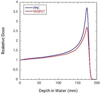
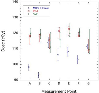
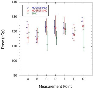
When in vivo proton dosimetry is performed with a metal-oxide semiconductor field-effect transistor (MOSFET) detector, the response of the detector depends strongly on the linear energy transfer. The present study reports a practical method to correct the MOSFET response for linear energy transfer dependence by using a simplified Monte Carlo dose calculation method (SMC). A depth-output curve for a mono-energetic proton beam in polyethylene was measured with the MOSFET detector. This curve was used to calculate MOSFET output distributions with the SMC (SMC(MOSFET)). The SMC(MOSFET) output value at an arbitrary point was compared with the value obtained by the conventional SMC(PPIC), which calculates proton dose distributions by using the depth-dose curve determined by a parallel-plate ionization chamber (PPIC). The ratio of the two values was used to calculate the correction factor of the MOSFET response at an arbitrary point. The dose obtained by the MOSFET detector was determined from the product of the correction factor and the MOSFET raw dose. When in vivo proton dosimetry was performed with the MOSFET detector in an anthropomorphic phantom, the corrected MOSFET doses agreed with the SMC(PPIC) results within the measurement error. To our knowledge, this is the first report of successful in vivo proton dosimetry with a MOSFET detector.
Figures
References
-
- Chuang CF, Verhey LJ, Xia P. Investigation of the use of MOSFET for clinical IMRT dosimetric verification. Med Phys. 2002; 29 (6): 1109–15. - PubMed
-
- Ramaseshan R, Kohli KS, Zhang TJ, et al. Performance characteristics of a microMOSFET as an in vivo dosimeter in radiation therapy. Phys Med Biol. 2004; 49 (17): 4031–48. - PubMed
-
- Ehringfeld C, Schmid S, Poljanc K, Kirisits C, Aiginger H, Georg D. Application of commercial MOSFET detectors for in vivo dosimetry in the therapeutic x‐ray range from 80 kV to 250 kV. Phys Med Biol. 2005; 50 (2): 289–303. - PubMed
-
- Kohno R, Hirano E, Nishio T, et al. Dosimetric evaluation of a MOSFET detector for clinical application in photon therapy. Radiol Phys Technol. 2008; 1 (1): 55–61. - PubMed
-
- Kohno R, Hirano E, Kitou S, et al. Evaluation of the usefulness of a MOSFET detector in an anthropomorphic phantom for 6‐MV photon beam. Radiol Phys Technol. 2010; 3 (2): 104–12. - PubMed
Publication types
MeSH terms
Substances
LinkOut - more resources
Full Text Sources
