. 2012 Jun;132(6):1605-14.
doi: 10.1038/jid.2012.32. Epub 2012 Mar 8.

Osteopontin in systemic sclerosis and its role in dermal fibrosis

Affiliations

Osteopontin in systemic sclerosis and its role in dermal fibrosis

Minghua Wu et al. J Invest Dermatol. 2012 Jun.

Abstract

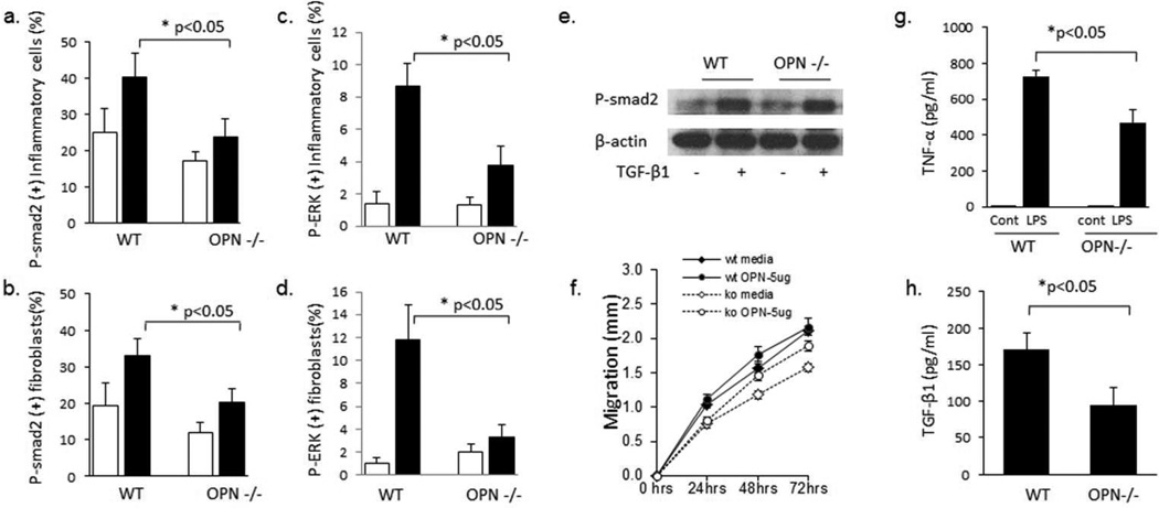
Osteopontin (OPN) is a matricellular protein with proinflammatory and profibrotic properties. Previous reports demonstrate a role for OPN in wound healing and pulmonary fibrosis. Here, we determined whether OPN levels are increased in a large cohort of patients with systemic sclerosis (SSc) and whether OPN contributes to the development of dermal fibrosis. The plasma OPN levels were increased in SSc patients, including patients with limited and diffuse disease, compared with healthy controls. Immunohistology demonstrated OPN on fibroblast-like and inflammatory cells in SSc skin and lesional skin from mice in the bleomycin (bleo)-induced dermal fibrosis model. OPN-deficient (OPN(-/-)) mice developed less dermal fibrosis compared with wild-type (WT) mice in the bleo-induced dermal fibrosis model. Additional in vivo studies have demonstrated that lesional skin from OPN(-/-)mice had fewer Mac-3-positive cells, fewer myofibroblasts, decreased transforming growth factor (TGF)-β and genes in the TGF-β pathway, and decreased numbers of cells expressing phosphorylated SMAD2 (pSMAD) and extracellular signal-regulated kinase. In vitro, OPN(-/-) dermal fibroblasts had decreased migratory capacity but similar phosphorylation of SMAD2 by TGF-β. Finally, TGF-β production by OPN-deficient macrophages was reduced compared with WT. These data demonstrate an important role for OPN in the development of dermal fibrosis and suggest that it may be a new therapeutic target in SSc.


Conflict of interest statement

Conflict of Interest: The authors declare no conflicts of interest.

Figures

Figure 1
Figure 1. Osteopontin expression in SSc patients and Fibrotic mouse dermis
(A) Plasma levels of OPN are increased in SSc patients overall and in limited and diffuse SSc compared to controls. Data presented as mean±SEM. (B) Plasma levels of OPN are increased in SSc-associated antibody (ACA, ATA, ARA) subsets of patients compared to controls. (C–H) IHC analyses with anti-OPN antibody demonstrates increased OPN immunoreactivity in skin from SSc patients (F–H) compared to controls (C–E). Scale bar is 200 µm (c,f), 50 µm (d, e) and 20 µm (e, h). (I) Increased number of fibroblasts (spindle shaped cells) and inflammatory cells (round cells) with OPN immunoreactivity were observed in control (open bars) compared to SSc skin (black bars). (J) qRTPCR analysis of total RNA from lesional skin of wild type mice received daily injection of bleo or PBS for 28 days demonstrates increased expression of OPN after bleo injection. Data presented as mean ± SEM of duplicated determinations from 10 mice per group. *p<0.05. (K) IHC analysis of bleo injected mouse skin demonstrated increased OPN expression compared to PBS injected skin. Scale bar is 200 µm in panel 1 and 3, and 50 µm in panel 4.
Figure 2
Figure 2. OPN−/− mice have reduced skin fibrosis in the bleomycin induced dermal fibrosis model
OPN−/− and wild type mice received daily s.c. injections of PBS or bleomycin for 28 days, and lesional skin was examined. (A) H&E stain and Masson’s trichrome (MT). Scale bar = 200 µm. (B) Quantification of dermal thickness. The results represent the mean±SEM, 15 mice/group. Open bars, PBS; closed bars, bleomycin. *p<0.001. (C) Soluble collagen was quantified by Sircol colorimetric assay (Data presented as mean ± SEM, from 15 mice/group. Open bars, PBS; closed bars, bleomycin. *p<0.05). (D) Total RNA from lesional skin was analyzed for Col1a1 mRNA by qRTPCR. Data presented as mean ± SEM of duplicate determinations from 6 mice/group, *p<0.05.
Figure 3
Figure 3. OPN−/− mice have decrease dermal inflammation
(A) IHC with anti-MAC-3 antibody was performed on lesional skin from mice injected with PBS or bleo The number of Mac-3 positive macrophages was quantified in 6 high powered fields per mouse (4 mice per group, **p<0.01). Inflammatory cytokine mRNA level were decreased in OPN−/− mice injected with bleomycin compared to wild type mice (b. CCL-2, c. IL-6; 6 mice per group, *p<0.05.) Data presented as mean ± SEM. Open bars, PBS; closed bars, bleomycin.
Figure 4
Figure 4. OPN−/− mice have decreased TGFβ and downstream pathways
(A) IHC with anti-SMA antibody was performed on lesional skin from mice injected with PBS or bleo. IHC with anti-SMA antibody demonstrated decreased myofibroblasts in OPN−/− mice Data presented as mean ± SEM of triplicate determinations in at least 6 microscopic fields, four mice/group. Open bars, PBS; closed bars, bleomycin. *p=0.0076. Total mRNA from lesional skin of OPN−/− mice injected with bleo demonstrated decreased levels of (B) SMA, (C) TGFβ, (D) CCN2, and (E) PAI-1.Data presented as mean ± SEM of duplicate determinations from 6 mice/group, *p<0.05.
Figure 5
Figure 5. OPN−/− mice have decreased phosphorylated SMAD2 in vivo but not in vitro
IHC with anti-pSMAD2 antibody (A,B) or pERK antibody (C,D) was performed on lesional skin from mice injected with PBS or bleo. OPN−/− mice had decrease numbers of pSMAD2 inflammatory cells (A) and fibroblasts (B) after bleo injection. OPN−/− mice also had decrease numbers of pERK inflammatory cells (C) and fibroblasts (D) after bleo injection. Data presented as mean ± SEM in at least 6 microscopic fields, 4 mice per group. In contrast, in vitro stimulation of dermal fibroblasts with TGFb resulted in similar levels of SMAD2 phosphorylation in wild type and OPN−/− cells (E). (F) OPN−/− dermal fibroblasts had less migratory capacity in the scratch wound assay compared to wild type dermal fibroblasts which is partially restored by 5 µg/ml of OPN (representative of n=3 WT and 3 OPN−/−). (G,H) Bone marrow derived macrophages OPN−/− mice produced less TNFα (G) and TGFβ (H) relative to WT macrophages (WT n=4, OPN−/− n=4). Data presented as mean ± SEM.

References

    1. Abraham DJ, Varga J. Scleroderma: from cell and molecular mechanisms to disease models. Trends Immunol. 2005;26:587–595. - PubMed
    1. Agarwal SK, Wu M, Livingston CK, Parks DH, Mayes MD, Arnett FC, et al. Toll-like receptor 3 upregulation by type I interferon in healthy and scleroderma dermal fibroblasts. Arthritis Res Ther. 2011;13:R3. - PMC - PubMed
    1. Anborgh PH, Mutrie JC, Tuck AB, Chambers AF. Pre- and post-translational regulation of osteopontin in cancer. J Cell Commun Signal. 2011;5:111–122. - PMC - PubMed
    1. Arnett FC. Is scleroderma an autoantibody mediated disease? Curr Opin Rheumatol. 2006;18:579–581. - PubMed
    1. Ashkar S, Weber GF, Panoutsakopoulou V, Sanchirico ME, Jansson M, Zawaideh S, et al. Eta-1 (osteopontin): an early component of type-1 (cell-mediated) immunity. Science. 2000;287:860–864. - PubMed

Publication types

MeSH terms