

Combined use of laser capture microdissection and cDNA microarray analysis identifies locally expressed disease-related genes in focal regions of psoriasis vulgaris skin lesions
- PMID: 22402443
- PMCID: PMC3352975
- DOI: 10.1038/jid.2012.33
Combined use of laser capture microdissection and cDNA microarray analysis identifies locally expressed disease-related genes in focal regions of psoriasis vulgaris skin lesions
Abstract
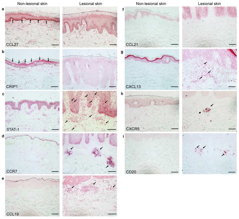
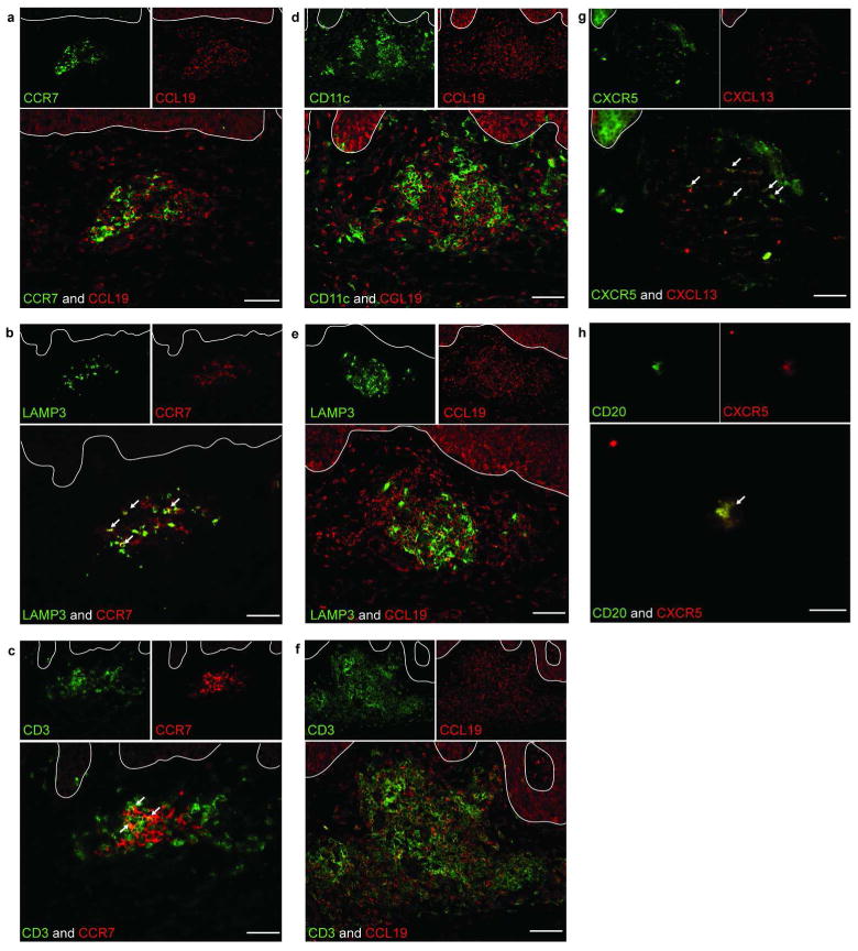
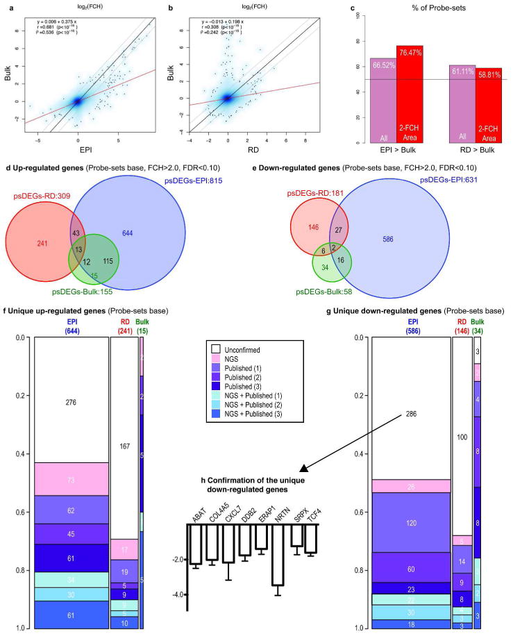
Psoriasis vulgaris is a complex disease characterized by alterations in growth and differentiation of epidermal keratinocytes, as well as a marked increase in leukocyte populations. Lesions are known to contain alterations in messenger RNAs encoding more than 1,000 products, but only a very small number of these transcripts has been localized to specific cell types or skin regions. In this study, we used laser capture microdissection (LCM) and gene array analysis to study the gene expression of cells in lesional epidermis (EPI) and dermis, compared with the corresponding non-lesional regions. Using this approach, we detected >1,800 differentially expressed gene products in the EPI or dermis of psoriasis lesions. These results established sets of genes that are differentially expressed between epidermal and dermal compartments, as well as between non-lesional and lesional psoriasis skin. One of our findings involved the local production of CCL19, a lymphoid-organizing chemokine, and its receptor CCR7 in psoriatic dermal aggregates, along with the presence of gene products LAMP3/DC-LAMP and CD83, which typify mature dendritic cells (DCs). Gene expression patterns obtained with LCM and microarray analysis along with T-cell and DC detection by immune staining suggest a possible mechanism for lymphoid organization via CCL19/CCR7 in diseased skin.
Conflict of interest statement
Figures
Comment in
-
Capturing the finer points of gene expression in psoriasis: beaming in on the CCL19/CCR7 axis.J Invest Dermatol. 2012 Jun;132(6):1535-8. doi: 10.1038/jid.2012.134. J Invest Dermatol. 2012. PMID: 22584500 Free PMC article.
References
-
- Bowcock AM, Shannon W, Du F, et al. Insights into psoriasis and other inflammatory diseases from large-scale gene expression studies. Hum Mol Genet. 2001;10:1793–1805. - PubMed
-
- Clark RA, Chong B, Mirchandani N, et al. The vast majority of CLA+ T cells are resident in normal skin. J Immunol. 2006;176:4431–4439. - PubMed
-
- Clemmesen A, Thomassen M, Clemmesen O, et al. Extraction of high-quality epidermal RNA after ammonium thiocyanate-induced dermo-epidermal separation of 4 mm human skin biopsies. Exp Dermatol. 2009;18:979–984. - PubMed
Publication types
MeSH terms
Grants and funding
LinkOut - more resources
Full Text Sources
Other Literature Sources
Medical
Molecular Biology Databases
