. 2012 Apr 3;109(14):5277-82.
doi: 10.1073/pnas.1114487109. Epub 2012 Mar 7.

Control of protein signaling using a computationally designed GTPase/GEF orthogonal pair

Affiliations

Control of protein signaling using a computationally designed GTPase/GEF orthogonal pair

Gregory T Kapp et al. Proc Natl Acad Sci U S A. .

Abstract

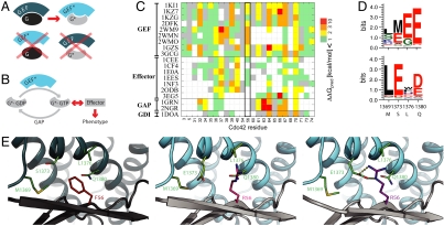
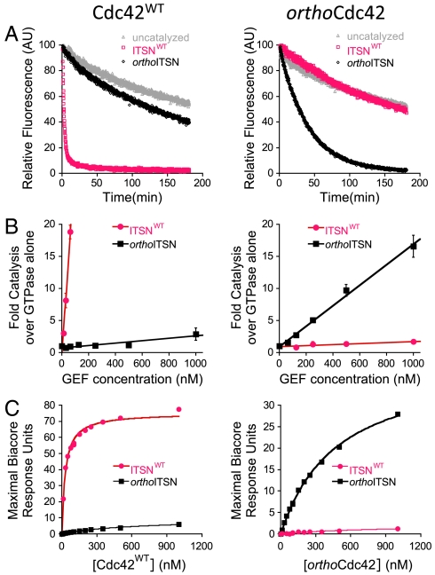
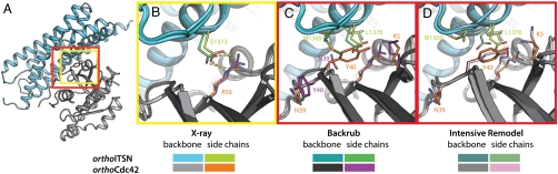
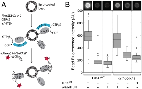
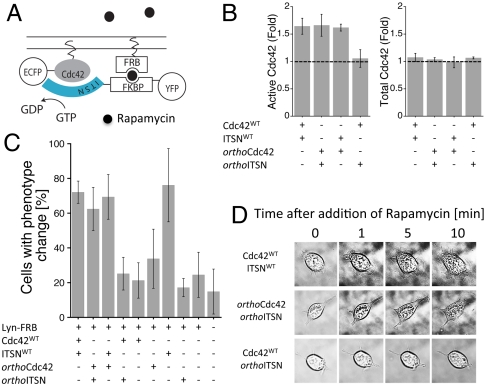
Signaling pathways depend on regulatory protein-protein interactions; controlling these interactions in cells has important applications for reengineering biological functions. As many regulatory proteins are modular, considerable progress in engineering signaling circuits has been made by recombining commonly occurring domains. Our ability to predictably engineer cellular functions, however, is constrained by complex crosstalk observed in naturally occurring domains. Here we demonstrate a strategy for improving and simplifying protein network engineering: using computational design to create orthogonal (non-crossreacting) protein-protein interfaces. We validated the design of the interface between a key signaling protein, the GTPase Cdc42, and its activator, Intersectin, biochemically and by solving the crystal structure of the engineered complex. The designed GTPase (orthoCdc42) is activated exclusively by its engineered cognate partner (orthoIntersectin), but maintains the ability to interface with other GTPase signaling circuit components in vitro. In mammalian cells, orthoCdc42 activity can be regulated by orthoIntersectin, but not wild-type Intersectin, showing that the designed interaction can trigger complex processes. Computational design of protein interfaces thus promises to provide specific components that facilitate the predictable engineering of cellular functions.


Conflict of interest statement

The authors declare no conflict of interest.

Figures

Fig. 1.
Fig. 1.
Strategy for computational design of an orthogonal signaling interaction. (A) Schematic representation of design requirements for orthogonality: the interface between the GTPase Cdc42 (G) and ITSN (GEF) is modified to generate a pair G*/GEF* with new specificity. (B) Simplified schematic representation of the core GTPase signaling circuit to define the design requirements for a functional G*/GEF* pair that interfaces correctly with other cellular components (GAP and effector proteins that are required for phenotypic output). (C) Computational alanine scanning. Shown are the estimated effects on binding energy of replacing each residue in the Cdc42/ITSN interface (PDB code 1KI1) with alanine in the context of 19 co-complex structures of Cdc42 with partner proteins (white indicates residues not in the interface in the respective structure). Altering position F56 of Cdc42 mainly affects interaction with GEFs. (D) Comparison of fixed backbone (top) and flexible backbone (bottom) computational design predictions for four residues in ITSN (wild-type residues are indicated on the x axis) in the vicinity of position 56 of Cdc42 for a F56R mutation. (E) Model of designed orthoCdc42/orthoITSN interface from fixed (middle) and flexible (right) backbone modeling compared to the wild-type complex (left). Gray: Cdc42; Teal: ITSN; shown in sticks are the five designed interface residues. Small backbone changes modeled by backrub motions (0.53 Å Cα rmsd) allowed the sidechains of R56 and E1373 to adopt conformations that can form hydrogen bonds (dashed lines).
Fig. 2.
Fig. 2.
The designed interaction is orthogonal in vitro. In (A)–(C), Cdc42WT is shown on the left and orthoCdc42 on the right. Pink: data for ITSNWT; black: data for orthoITSN. (A) Catalysis of nucleotide exchange by ITSNWT and orthoITSN, monitored by dissociation of fluorescent mant-GDP from Cdc42WT and orthoCdc42. Gray: intrinsic exchange in Cdc42 in the absence of any ITSN. (B) Catalysis of nucleotide exchange from initial rates of mant-GDP association at varying GEF concentrations. Data represent averages and standard deviations from at least three experiments. (C). Binding affinity monitored by Surface Plasmon Resonance equilibrium analysis (SI Appendix, Fig. S4).
Fig. 3.
Fig. 3.
The crystal structure of the orthoCdc42/orthoITSN complex confirms the designed interaction, but also highlights requirements for advanced flexible-backbone remodeling protocols. (A) Overview of the structure of the designed complex between orthoCdc42 (gray) and the orthoITSN DH domain (teal). Boxes highlight the location of the designed site near the center of the protein-protein interface (yellow) as well as the area of backbone and side-chain rearrangements (red), magnified in (B–D). Sidechain and backbone colors are as indicated in the figure. (B) Comparison of the R56-E1373 interaction in the backrub flexible-backbone computational model (as in Fig. 1E, right) and in the crystal structure of the designed orthoCdc42/orthoITSN complex. Dashed lines represent hydrogen bonds. (C, D) Comparison of the network of residues surrounding the designed site that were rearranged to accommodate the mutations, as predicted by the backrub model (C) and the intensive remodeling protocol (D, details in SI Appendix, Results) vs. their observed position in the crystal structure of the designed complex. The remodeling protocol (D) was able to capture both sidechain and backbone conformational changes in the crystal structure of orthoCdc42/orthoITSN that were missed by the initial backrub predictions (C).
Fig. 4.
Fig. 4.
The designed orthoCdc42/orthoITSN interaction mediates specific GTPase activation and effector binding in an in vitro reconstituted system. Alexa 594 labeled N-WASP (residues 137–502) translocation to a lipid-coated glass bead is specifically increased in the presence of a cognate interaction between Cdc42 and ITSN. (A) Schematic illustrating the assay and the order of addition of the components. (B) The total fluorescence intensity of individual beads relative to the background was measured, and the distributions of the fluorescence intensities from multiple beads (n > 23 for each condition) are shown in box plot representation. Boxes enclose the first and third quartile of the distribution and display a line at the median; whiskers extend outward no more than 1.5 times the size of the box and data points outside this range are drawn individually. A representative bead image is shown above each condition.
Fig. 5.
Fig. 5.
The orthoCdc42/orthoITSN pair is functional in mammalian cells. (A) Schematic representation of the cell-based assay using a Rapamycin-based recruitment system (FRB, FKPB) to colocalize fluorescently tagged GTPase and GEF constructs at the membrane. (B) Fold increase in active Cdc42 (comparing samples with and without addition of Rapamycin for 60 s) from lysed NIH 3T3 cells measured with a G-LISA assay (left). The total Cdc42 loaded in the G-LISA assay was determined by an ELISA assay, and is also shown in fold change, again comparing samples with and without Ramamycin addition (right). All samples had Lyn-FRB transfected. Error bars represent the standard deviation of three experiments. (C) Percentage of NIH 3T3 cells that showed morphological changes (filopodia/lamellipodia) after addition of Rapamycin, determined by live cell microscopy. All samples had Lyn-FRB transfected. Error bars represent the standard deviation of three experiments. The total numbers of counted cells for each condition, from left to right, are: 103, 111, 133, 120, 57, 62, 55, 50, 71, and 84. (D) Representative images of cell morphological changes upon Rapamycin addition.

Comment in

References

    1. Elowitz MB, Leibler S. A synthetic oscillatory network of transcriptional regulators. Nature. 2000;403:335–338. - PubMed
    1. Gardner TS, Cantor CR, Collins JJ. Construction of a genetic toggle switch in Escherichia coli. Nature. 2000;403:339–342. - PubMed
    1. Sprinzak D, Elowitz MB. Reconstruction of genetic circuits. Nature. 2005;438:443–448. - PubMed
    1. Yeh BJ, Rutigliano RJ, Deb A, Bar-Sagi D, Lim WA. Rewiring cellular morphology pathways with synthetic guanine nucleotide exchange factors. Nature. 2007;447:596–600. - PubMed
    1. Bashor CJ, Helman NC, Yan S, Lim WA. Using engineered scaffold interactions to reshape MAP kinase pathway signaling dynamics. Science. 2008;319:1539–1543. - PubMed

Publication types

Associated data