. 2012 Mar 27;109(13):5010-5.
doi: 10.1073/pnas.1114931109. Epub 2012 Mar 7.

Intestinal CX3C chemokine receptor 1(high) (CX3CR1(high)) myeloid cells prevent T-cell-dependent colitis

Affiliations

Intestinal CX3C chemokine receptor 1(high) (CX3CR1(high)) myeloid cells prevent T-cell-dependent colitis

Hisako Kayama et al. Proc Natl Acad Sci U S A. .

Abstract

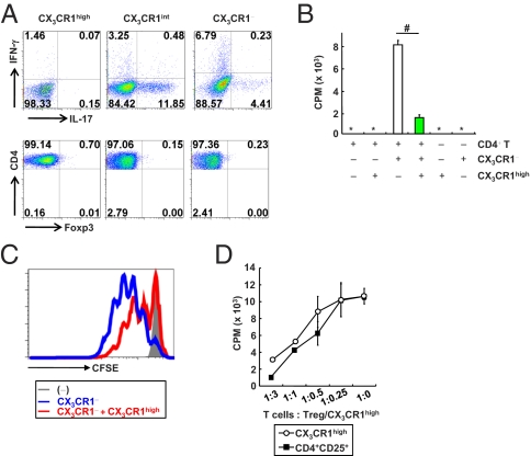
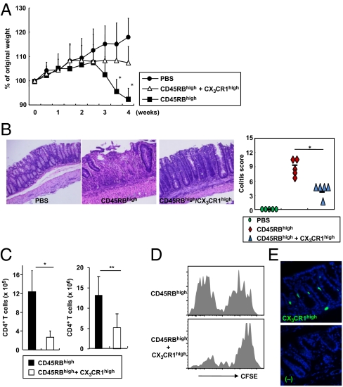
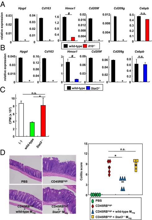
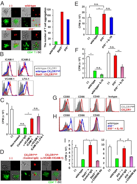
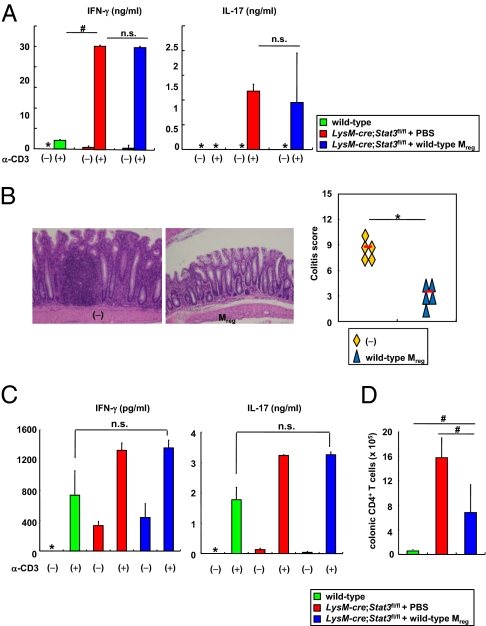
Adequate activation of CD4(+) T lymphocytes is essential for host defense against invading pathogens; however, exaggerated activity of effector CD4(+) T cells induces tissue damage, leading to inflammatory disorders such as inflammatory bowel diseases. Several unique subsets of intestinal innate immune cells have been identified. However, the direct involvement of innate immune cell subsets in the suppression of T-cell-dependent intestinal inflammation is poorly understood. Here, we report that intestinal CX(3)C chemokine receptor 1(high) (CX(3)CR1(high)) CD11b(+) CD11c(+) cells are responsible for prevention of intestinal inflammation through inhibition of T-cell responses. These cells inhibit CD4(+) T-cell proliferation in a cell contact-dependent manner and prevent T-cell-dependent colitis. The suppressive activity is abrogated in the absence of the IL-10/Stat3 pathway. These cells inhibit T-cell proliferation by two steps. Initially, CX(3)CR1(high) CD11b(+) CD11c(+) cells preferentially interact with T cells through highly expressed intercellular adhesion molecule-1/vascular cell adhesion molecule-1; then, they fail to activate T cells because of defective expression of CD80/CD86. The IL-10/Stat3 pathway mediates the reduction of CD80/CD86 expression. Transfer of wild-type CX(3)CR1(high) CD11b(+) CD11c(+) cells prevents development of colitis in myeloid-specific Stat3-deficient mice. Thus, these cells are regulatory myeloid cells that are responsible for maintaining intestinal homeostasis.


Conflict of interest statement

The authors declare no conflict of interest.

Figures

Fig. 1.
Fig. 1.
CX3CR1high CD11b+ CD11c+ cells in the intestinal lamina propria suppress T-cell proliferation. (A) Flow cytometric plots of IL-17-, IFN-γ-, or Foxp3-expressing CD4+ T cells cocultured with the indicated cells for 72 h. (B) [3H]thymidine uptake of CD4+ T cells cultured with the indicated cells. #P < 0.022. (C) The fluorescence intensity of CFSE-labeled CD4+ T cells cultured with the indicated cells at a ratio 1:1:1 for 72 h. (D) [3H]thymidine uptake by CD4+ T cells cocultured with CX3CR1 DCs in the presence of increasing ratios of splenic CD4+ CD25+ Treg cells (closed rectangle) or colonic CX3CR1high cells (open circle). All data are representative of two independent experiments (means ± SD of duplicate well measurements).
Fig. 2.
Fig. 2.
CX3CR1high myeloid cells alleviate T-cell-dependent intestinal inflammation. (A) SCID mice were injected i.p. with 3 × 10⁵ CD45RBhigh CD4+ T cells or PBS (closed circles). After 2 h, 3 × 105 CX3CR1high cells were transferred (open triangles) or not (closed rectangles). Body weight change was monitored and is presented relative to initial body weight. *P < 0.005 (n = 8 per group). (B) Hematoxylin and eosin staining of colon sections at 4 wk after the transfer described in A (Left) and the colitis score (Right). *P < 0.0012. (Original magnification, 200×.) (C) Numbers of large intestinal lamina propria CD4+ T cells at 2 wk (n = 4 per group) (Left) and 4 wk (n = 5 per group) (Right) after transfer. *P < 0.02; **P < 0.015. (D) CD45RBhigh T cells (3 × 10⁵) were labeled with CFSE and transferred into Rag2−/− mice with or without 3 × 105 CX3CR1high cells. After 12 d, CFSE dilution in colonic CD4+ T cells was analyzed. (E) Cryosection of the colon from a SCID mouse at 3 d after i.v. injection of CFSE-labeled CX3CR1high cells. (Original magnification, 200×.) Data are representative of three independent experiments (D and E).
Fig. 3.
Fig. 3.
Defective activity of Stat3−/− Mreg cells. (A and B) Expression of Hpgd, Cd163, Hmox1, CD209f, CD209g, and Cebpb mRNA in CX3CR1high Mreg cells from wild-type, Il10−/−, and LysM-cre; Stat3fl/fl mice. Data are presentative of two independent experiments (means ± SD of at least triplicate PCRs on the identical sample). *, not detected; #P < 0.025. (C) [3H]thymidine uptake by CD4+ T cells cultured with CX3CR1 DCs in the presence of wild-type or Stat3−/− CX3CR1high Mreg cells. Data are representative of four independent experiments (means ± SD of triplicate well measurements). *P < 0.047. (D) Hematoxylin and eosin staining of colon sections of Rag2−/− mice given 3 × 10⁵ CD45RBhigh CD4+ T cells with 3 × 10⁵ Mreg cells from wild-type or LysM-cre; Stat3fl/fl mice (Left) and colitis score (Right). (Original magnification, 200×.) *P < 0.025 (n = 5 per group).
Fig. 4.
Fig. 4.
Two-step mechanism for suppression of T-cell growth by Mreg cells. (A) Green dye-labeled CD4+ T cells were cultured with nonlabeled CX3CR1 DCs and red dye-labeled Mreg cells from wild-type, LysM-cre; Stat3fl/fl, or Il10−/− mice (Left). The number of T-cell aggregation in twenty fields (Right). (Original magnification, 100×.) (B) Expression of adhesion molecules on the indicated cells from wild-type and LysM-cre; Stat3fl/fl mice. (C) CD4+ T cells and wild-type CX3CR1 DCs were cocultured with Mreg cells pretreated with the indicated blocking Abs or control Ig for evaluation of T-cell proliferation. *P < 0.012. (D) Red dye-labeled CX3CR1high Mreg cells were treated with blocking Abs to ICAM-1 and VCAM-1 then added to the mixture of green dye-labeled CD4+ T cells and nonstained CX3CR1 DCs. (Original magnification, 100×.) (E) IL-10 (100 ng/mL) was added to the coculture of CD4+ T cells, wild-type DCs, and Il10−/− Mreg cells. Then, T-cell proliferation was measured. *P < 0.02. (F) Wild-type and Il10−/− Mreg cells were preincubated with or without 100 ng/mL IL-10 for 72 h. Then, the cells were analyzed for the suppressive activity of T-cell proliferation. *P < 0.016; **P < 0.034. (G) Surface expression of CD80, CD86, CD40, and MHC class II on CX3CR1high Mreg cells and CX3CR1 DCs. (H) Expression of CD80, CD86, and CD40 on Mreg cells from wild-type and Il10−/− mice cultured for 48 h with or without 100 ng/mL IL-10. (I) Stat3−/− and Il10−/− Mreg cells were pretreated with the indicated blocking Abs. Then, Mreg cells were cultured with CD4+ T cells and wild-type CX3CR1 DCs, and T-cell proliferation was measured. *P < 0.025; #P < 0.045. All data are representative of at least two independent experiments (mean values ± SD of triplicate well measurements).
Fig. 5.
Fig. 5.
Defective Mreg cell function leads to development of colitis. (A, C, and D) LysM-cre; Stat3fl/fl mice were transferred i.p. with 7 × 10⁴ wild-type CX3CR1high Mreg cells at 4 and 6 wk of age. At 2 wk after the last transfer, splenic (A) or colonic lamina propria (C) CD4+ T cells were analyzed for production of IFN-γ and IL-17A. *, not detected; #P < 0.008. Total number of CD4+ T cells in the colonic lamina propria was analyzed (D). #P < 0.0079. Data are from two independent experiments with four mice per group. (B) Hematoxylin and eosin staining of colon sections at 2 wk after the last transfer (Left) and the colitis score (Right). (Original magnification, 200×.) *P < 0.0002.

References

    1. Bouma G, Strober W. The immunological and genetic basis of inflammatory bowel disease. Nat Rev Immunol. 2003;3:521–533. - PubMed
    1. Xavier RJ, Podolsky DK. Unravelling the pathogenesis of inflammatory bowel disease. Nature. 2007;448:427–434. - PubMed
    1. Strober W, Fuss I, Mannon P. The fundamental basis of inflammatory bowel disease. J Clin Invest. 2007;117:514–521. - PMC - PubMed
    1. Wing K, Sakaguchi S. Regulatory T cells exert checks and balances on self tolerance and autoimmunity. Nat Immunol. 2010;11:7–13. - PubMed
    1. Sakaguchi S, Wing K, Onishi Y, Prieto-Martin P, Yamaguchi T. Regulatory T cells: How do they suppress immune responses? Int Immunol. 2009;21:1105–1111. - PubMed

Publication types

MeSH terms