. 2012 Apr 26;119(17):3997-4008.
doi: 10.1182/blood-2011-11-392985. Epub 2012 Mar 8.

Functional STAT3 deficiency compromises the generation of human T follicular helper cells

Affiliations

Functional STAT3 deficiency compromises the generation of human T follicular helper cells

Cindy S Ma et al. Blood. .

Abstract

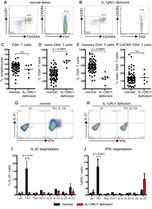
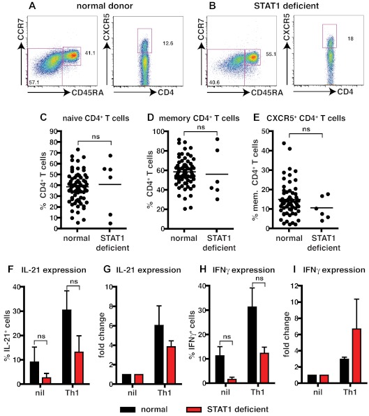
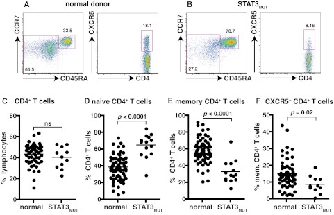
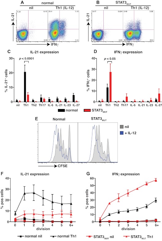
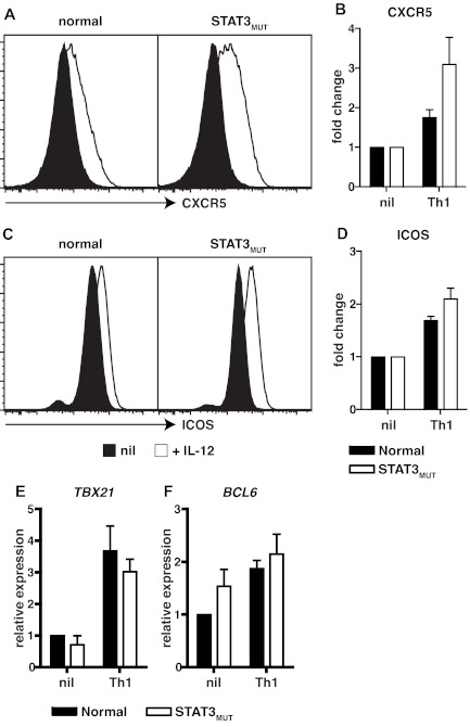
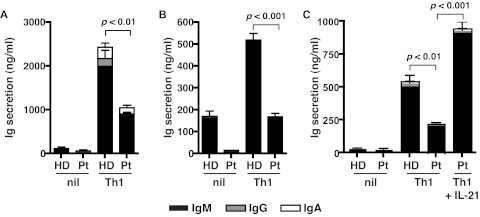
T follicular helper (Tfh) cells are critical for providing the necessary signals to induce differentiation of B cells into memory and Ab-secreting cells. Accordingly, it is important to identify the molecular requirements for Tfh cell development and function. We previously found that IL-12 mediates the differentiation of human CD4(+) T cells to the Tfh lineage, because IL-12 induces naive human CD4(+) T cells to acquire expression of IL-21, BCL6, ICOS, and CXCR5, which typify Tfh cells. We have now examined CD4(+) T cells from patients deficient in IL-12Rβ1, TYK2, STAT1, and STAT3 to further explore the pathways involved in human Tfh cell differentiation. Although STAT1 was dispensable, mutations in IL12RB1, TYK2, or STAT3 compromised IL-12-induced expression of IL-21 by human CD4(+) T cells. Defective expression of IL-21 by STAT3-deficient CD4(+) T cells resulted in diminished B-cell helper activity in vitro. Importantly, mutations in STAT3, but not IL12RB1 or TYK2, also reduced Tfh cell generation in vivo, evidenced by decreased circulating CD4(+)CXCR5(+) T cells. These results highlight the nonredundant role of STAT3 in human Tfh cell differentiation and suggest that defective Tfh cell development and/or function contributes to the humoral defects observed in STAT3-deficient patients.


Figures

Figure 1
Figure 1
Naive CD4+ T cells deficient for IL-12Rβ1 fail to differentiate into IL-21–expressing cells in response to IL-12. (A-F) The frequency of total CD4+ T cells, and naive (CD45RA+CCR7+), memory (CD45RA+CCR7−/+), and CXCR5+CD45RA CD4+ T cells, in PBMCs was determined for healthy donors and patients deficient for IL-12Rβ1. (A-B) Representative dot plots from 1 donor and 1 patient. (C-F) The frequency of (C) total, (D) naive (CD45RA+CCR7+), (E) memory (CD45RA+CCR7−/+), and (F) CXCR5+CD45RA CD4+ T cells from all healthy donors (total CD4+ T cells, n = 54; naive CD4+ T cells, n = 70; memory CD4+ T cells, n = 70; CXCR5+CD45RA CD4+ T cells, n = 61) and patients deficient for IL-12Rβ1 examined (n = 6). (G-J) Naive CD4+ T cells isolated from healthy donors (n = 5) and patients deficient for IL-12Rβ1 (n = 5) were cultured for 5 days under neutral (nil); polarizing Th1, Th2, or Th17 conditions; or in the presence of IL-6, IL-21, IL-23, or IL-27, and intracellular expression of IL-21 and IFNγ were then determined. (G-H) Representative dot plots of IL-21 and IFNγ expression by activated naive CD4+ T cells from 1 donor and 1 patient deficient for IL-12Rβ1. (I-J) Percentage of activated normal and IL-12Rβ1–deficient naive CD4+ T cells induced to express (I) IL-21 or (J) IFNγ in response to the indicated culture. The values represent the mean ± SEM.
Figure 2
Figure 2
STAT1 is dispensable for IL-12–induced expression of IL-21 in human CD4+ T cells. (A-E) The frequency of naive (CD45RA+CCR7+), memory (CD45RA+CCR7−/+), and CXCR5+CD45RA CD4+ T cells in PBMCs was determined for healthy donors and STAT1-deficient patients. (A-B) Representative dot plots from 1 donor and 1 STAT1-deficient patient. (C-E) The frequency of (C) naive (CD45RA+CCR7+), (D) memory (CD45RA+CCR7−/+), and (E) CXCR5+CD45RA CD4+ T cells from all healthy donors (total CD4+ T cells, n = 54; naive CD4+ T cells, n = 70; memory CD4+ T cells, n = 70; CXCR5+CD45RA CD4+ T cells, n = 61) and STAT1-deficient patients (n = 6) was examined. (F-I) Total CD4+ T cells isolated from healthy donors and STAT1-deficient patients (n = 3) were cultured for 5 days under neutral (nil) or Th1-polarizing (ie, IL-12) conditions, and expression of intracellular IL-21 (F-G) and IFNγ (H-I) was then determined. The graphs in panels F and H show the frequency of cytokine-positive cells; those in panels G and I depict cytokine expression after Th1 polarization as fold-increase relative to the nil culture in each experiment. The values represent the mean ± SEM (n = 3).
Figure 3
Figure 3
Mutations in STAT3 compromise the generation of CD4+ memory and CXCR5+CD45RA Tfh cells. (A-B) The frequency of naive (CD45RA+CCR7+), memory (CD45RA+CCR7−/+), and CXCR5+CD45RA CD4+ T cells in PBMCs was determined for healthy donors and STAT3MUT patients. (A-B) Representative dot plots from 1 donor and 1 patient. (C-F) The frequency of (C) total CD4+ T cells, (D) naive (CD45RA+CCR7+), (E) memory (CD45RA+CCR7−/+), and (F) CXCR5+CD45RA CD4+ T cells from all healthy donors (total CD4+ T cells, n = 54; naive CD4+ T cells, n = 70; memory CD4+ T cells, n = 70; CXCR5+CD45RA CD4+ T cells, n = 61) and STAT3MUT patients (n = 14) was examined.
Figure 4
Figure 4
STAT3 mutations impair IL-12–induced expression of IL-21 in naive CD4+ T cells. (A-D) Naive CD4+ T cells isolated from healthy donors (n = 8) and STAT3MUT patients (n = 9) were cultured for 5 days under neutral conditions (nil); polarizing Th1, Th2, or Th17 conditions; or in the presence of IL-6, IL-21, IL-23, or IL-27, after which time expression of intracellular IL-21 and IFNγ was determined. (A-B) Representative dot plots of cytokine expression by activated naive CD4+ T cells from 1 donor and 1 patient. (C-D) Percentage of activated normal and STAT3MUT naive CD4+ T cells induced to express (C) IL-21 or (D) IFNγ in response to the indicated culture. The values represent the mean ± SEM. (E-F) Naive CD4+ T cells isolated from healthy donors and STAT3MUT patients were labeled with CFSE and cultured with anti-CD2/CD3/CD28 Abs in the absence (nil) or presence of Th1 polarizing conditions (+IL-12). After 5 days cells were harvested, and (E) proliferation and expression of (F) IL-21 and (G) IFNγ in cells that had undergone different divisions were then determined. (F-G) The values represent the mean ± SEM (n = 3).
Figure 5
Figure 5
Induction of CXCR5, ICOS, and BCL6 by IL-12 in CD4+ T cells is independent of STAT3. (A-D) Naive CD4+ T cells isolated from healthy donors and STAT3MUT patients were cultured with anti-CD2/CD3/CD28 Abs in the absence (nil) or presence of Th1-polarizing conditions (+IL-12). After 4 days the cells were harvested, and surface expression of CXCR5 and ICOS was determined by flow cytometry and of TBX21 and BCL6 by qPCR. (A,C) representative histogram plots from 1 healthy donor and 1 STAT3MUT patient. Expression of (B) CXCR5 (n = 3), (D) ICOS (n = 3), (E) TBX21 (n = 4), and (F) BCL6 (n = 5) after Th1 polarization is presented as fold-increase compared with the nil culture in each experiment. (B,D-F) The graphs represent the mean ± SEM of the indicated number of experiments.
Figure 6
Figure 6
STAT3-deficient cells show impaired Tfh cell function in vitro. Naive CD4+ T cells isolated from healthy donors (HD) and STAT3MUT patients (Pt) were cultured under neutral (nil) or Th1 polarizing conditions. After 5 days, the cells were harvested and treated with mitomycin C before being cocultured with allogeneic naive B cells, in the absence or presence of exogenous IL-21, for an additional 7 days. After this time secretion of IgM, IgG, and IgA was determined. (A-B) The data were derived from experiments that used naive CD4+ T cells isolated from different STAT3MUT patients; (C) the data show the effect of exogenous IL-21 on the ability of STAT3MUT CD4+ T cells to provide B-cell help. Each graph represents the mean ± SEM of triplicate cultures; similar results were obtained in 5 (A-B) and 2 (C) experiments.

References

    1. Tangye SG, Deenick EK, Palendira U, Ma CS. T cell-B cell interactions in primary immunodeficiencies. Ann N Y Acad Sci. 2012;1250(1):1–13. - PubMed
    1. Breitfeld D, Ohl L, Kremmer E, et al. Follicular B helper T cells express CXC chemokine receptor 5, localize to B cell follicles, and support immunoglobulin production. J Exp Med. 2000;192(11):1545–1552. - PMC - PubMed
    1. Schaerli P, Willimann K, Lang AB, Lipp M, Loetscher P, Moser B. CXC chemokine receptor 5 expression defines follicular homing T cells with B cell helper function. J Exp Med. 2000;192(11):1553–1562. - PMC - PubMed
    1. Chtanova T, Tangye SG, Newton R, et al. T follicular helper cells express a distinctive transcriptional profile, reflecting their role as non-Th1/Th2 effector cells that provide help for B cells. J Immunol. 2004;173(1):68–78. - PubMed
    1. Yu D, Rao S, Tsai LM, et al. The transcriptional repressor Bcl-6 directs T follicular helper cell lineage commitment. Immunity. 2009;31(3):457–468. - PubMed

Publication types

MeSH terms