. 2012 May;112(10):1678-88.
doi: 10.1152/japplphysiol.00060.2012. Epub 2012 Mar 8.

Severe acute intermittent hypoxia elicits phrenic long-term facilitation by a novel adenosine-dependent mechanism

Affiliations

Severe acute intermittent hypoxia elicits phrenic long-term facilitation by a novel adenosine-dependent mechanism

Nicole L Nichols et al. J Appl Physiol (1985). 2012 May.

Abstract

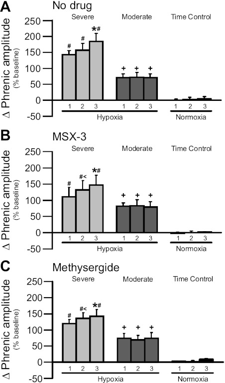
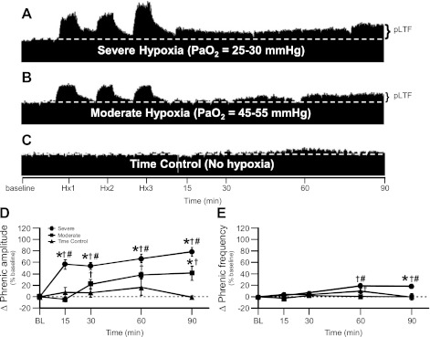
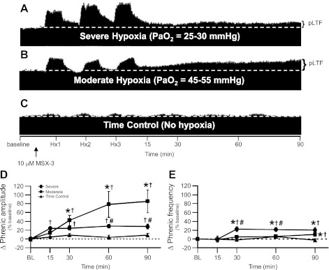
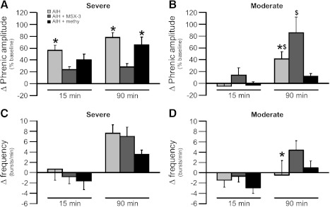
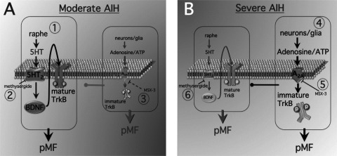
Acute intermittent hypoxia [AIH; 3, 5-min episodes; 35-45 mmHg arterial PO(2) (Pa(O(2)))] elicits serotonin-dependent phrenic long-term facilitation (pLTF), a form of phrenic motor facilitation (pMF) initiated by G(q) protein-coupled metabotropic 5-HT(2) receptors. An alternate pathway to pMF is induced by G(s) protein-coupled metabotropic receptors, including adenosine A(2A) receptors. AIH-induced pLTF is dominated by the serotonin-dependent pathway and is actually restrained via inhibition from the adenosine-dependent pathway. Here, we hypothesized that severe AIH shifts pLTF from a serotonin-dependent to an adenosine-dependent form of pMF. pLTF induced by severe (25-30 mmHg Pa(O(2))) and moderate (45-55 mmHg Pa(O(2))) AIH were compared in anesthetized rats, with and without intrathecal (C4) spinal A(2A) (MSX-3, 130 ng/kg, 12 μl) or 5-HT receptor antagonist (methysergide, 300 μg/kg, 15 μl) injections. During severe, but not moderate AIH, progressive augmentation of the phrenic response during hypoxic episodes was observed. Severe AIH (78% ± 8% 90 min post-AIH, n = 6) elicited greater pLTF vs. moderate AIH (41% ± 12%, n = 8; P < 0.05). MSX-3 (28% ± 6%; n = 6; P < 0.05) attenuated pLTF following severe AIH, but enhanced pLTF following moderate AIH (86% ± 26%; n = 8; P < 0.05). Methysergide abolished pLTF after moderate AIH (12% ± 5%; n = 6; P = 0.035), but had no effect after severe AIH (66 ± 13%; n = 5; P > 0.05). Thus severe AIH shifts pLTF from a serotonin-dependent to an adenosine-dependent mechanism; the adenosinergic pathway inhibits the serotonergic pathway following moderate AIH. Here we demonstrate a novel adenosine-dependent pathway to pLTF following severe AIH. Shifts in the mechanisms of respiratory plasticity provide the ventilatory control system greater flexibility as challenges that differ in severity are confronted.


Figures

Fig. 1.
Fig. 1.
Progressive augmentation of the short-term hypoxic phrenic response during severe (light gray bars) but not moderate (dark gray bars) hypoxic episodes or time control groups (normoxia; black bars) where 1, 2, and 3 represent hypoxic or normoxic episodes 1, 2, and 3. In A, the hypoxic response after no drug was administered and during severe acute intermittent hypoxia (AIH) progressively increases with each episode. [Asterisk (*) indicates 3 is significantly greater than 1 and 2]. Neither intrathecal MSX-3 (B) nor methysergide (C) alters this effect, demonstrating that progressive augmentation is independent of spinal serotonin or A2A receptor activation. Moderate AIH did not elicit progressive augmentation, similar to previous reports from our laboratory. There were no changes in time control experiments. Plus (+) indicates a greater response relative to time control experiments at equivalent times. (#) indicates that 1, 2, and 3 during moderate AIH and time control experiments are significantly lower than severe 1, 2, and 3, respectively. (<) indicates 2 is significantly higher than 1 in B and C. Values are means ± 1 SE; significance is P < 0.05.
Fig. 2.
Fig. 2.
Severe AIH elicits enhanced phrenic long-term facilitation (pLTF) vs. pLTF induced by moderate AIH. A–C: representative traces of compressed, integrated phrenic neurograms during severe and moderate AIH compared with rats treated as time controls and not exposed to AIH (white, dashed line in each trace indicates baseline). In A, rats exposed to severe AIH exhibit enhanced pLTF. In B, rats exposed to moderate AIH exhibit typical pLTF. In C, time control rats not exposed to AIH do not exhibit time-dependent changes in phrenic motor output. D: phrenic burst amplitude (percentage change from baseline) in severe (n = 6; circles) and moderate AIH (n = 8; squares) protocols compared with rats treated as time controls (n = 6; triangles) and not exposed to AIH. pLTF is significantly elevated following severe AIH at 15, 30, 60, and 90 min post-hypoxia compared with baseline (BL; †), rats treated with moderate AIH (#) and rats treated as time controls (*) (all P < 0.05). pLTF is significantly elevated following moderate AIH at 30, 60, and 90 min post-hypoxia compared with BL (†) and rats treated as time controls at 90 min post-hypoxia (*) (P < 0.05). E. Phrenic frequency (percentage change from baseline) in severe (circles) and moderate AIH (squares) protocols compared with rats treated as time controls (triangles) and not exposed to AIH, where phrenic frequency was scaled the same as phrenic burst amplitude to enable direct comparisons of magnitude. Minimal LTF in phrenic nerve burst frequency is seen with severe AIH at 60 and 90 min posthypoxia compared with BL (†), rats treated with moderate AIH (#) and only at 90 min post-hypoxia compared with rats treated as time controls (*) (all P < 0.05). Minimal LTF in phrenic nerve burst frequency is seen with moderate AIH only at 60 min posthypoxia compared with BL (†) (P < 0.05).
Fig. 3.
Fig. 3.
Severe, not moderate, AIH-induced pLTF is significantly attenuated by pretreatment with 10 μM MSX-3. A–C: representative traces of compressed, integrated phrenic neurograms during severe and moderate AIH compared with rats treated as time controls and not exposed to AIH after 10 μM MSX-3 spinal delivery (white, dashed line in each trace indicates baseline and black arrow indicates time of 10 μM MSX-3 delivery which was ∼5 min before the first hypoxic episode in each group). In A, rats exposed to severe AIH after spinal MSX-3 exhibit attenuated pLTF. In B, rats exposed to moderate AIH after spinal MSX-3 exhibit pronounced pLTF. In C, time control rats not exposed to AIH after spinal MSX-3 do not exhibit time-dependent changes in phrenic motor output. D: phrenic burst amplitude (percentage change from baseline) in severe (n = 6; circles) and moderate AIH (n = 8; squares) protocols compared with rats treated as time controls (n = 7; triangles) and not exposed to AIH after 10 μM MSX-3 spinal delivery. pLTF is significantly elevated by moderate AIH after 10 μM MSX-3 at 15, 30, 60, and 90 min posthypoxia compared with BL (†), and at 30, 60, and 90 min posthypoxia compared with rats treated as time controls (*) (all P < 0.05). pLTF following severe AIH after 10 μM MSX-3 is significantly attenuated compared with moderate AIH at 60 and 90 min posthypoxia (#) (P < 0.05) and is significantly increased at 30, 60, and 90 min posthypoxia compared with BL (†) (P < 0.05). E: phrenic frequency (percentage change from baseline) in severe (circles) and moderate AIH (squares) protocols compared with rats treated as time controls (triangles) and not exposed to AIH after 10 μM MSX-3 spinal delivery, where phrenic frequency was scaled the same as phrenic burst amplitude to enable direct comparisons of magnitude. Minimal LTF in phrenic nerve burst frequency is seen with severe AIH at 30, 60, and 90 min posthypoxia compared with BL (†) and rats treated as time controls (*), but only at 30 and 60 min posthypoxia compared with moderate AIH (#) (all P < 0.05). Minimal LTF in phrenic nerve burst frequency is seen with moderate AIH at 90 min posthypoxia compared with BL (†) and rats treated as time controls (*) (P < 0.05).
Fig. 4.
Fig. 4.
Moderate, not severe, AIH-induced pLTF is significantly inhibited by 20 mM methysergide. A–C: representative traces of compressed, integrated phrenic neurograms during severe and moderate AIH compared with rats treated as time controls and not exposed to AIH after 20 mM methysergide spinal delivery (white, dashed line in each trace indicates baseline and black arrow indicates time of 20 mM methysergide delivery which was ∼10 min before the first hypoxic episode in each group). In A, enhanced pLTF (after severe AIH) is not altered in rats pretreated with spinal methysergide. In B, rats exposed to moderate AIH after spinal methysergide delivery exhibit abolished pLTF. In C, time control rats not exposed to AIH after spinal methysergide delivery do not exhibit time-dependent changes in phrenic motor output. D: phrenic burst amplitude (percentage change from baseline) in severe (n = 5; circles) and moderate AIH (n = 6; squares) protocols compared with rats treated as time controls (n = 5; triangles) and not exposed to AIH after 20 mM methysergide spinal delivery. pLTF is significantly increased by severe AIH after 20 mM methysergide delivery at 15, 30, 60, and 90 min posthypoxia compared with BL (†), at 15, 60 and 90 min posthypoxia compared with rats treated with moderate AIH (#) and at 60 and 90 min posthypoxia compared with rats treated as time controls (*) (all P < 0.05). Notice that phrenic amplitude in rats treated with moderate AIH after 20 mM methysergide is not significantly different from BL or from rats treated as time controls; thus moderate AIH induced pLTF is inhibited by 20 mM methysergide. E: phrenic frequency (percentage change from baseline) in severe (circles) and moderate AIH (squares) protocols compared with rats treated as time controls (triangles) and not exposed to AIH after 20 mM methysergide spinal delivery, where phrenic frequency was scaled the same as phrenic burst amplitude to enable direct comparisons of magnitude. Minimal LTF in phrenic nerve burst frequency is only seen with severe AIH at 60 min posthypoxia compared with BL (†), (P < 0.05).
Fig. 5.
Fig. 5.
Direct comparison of the change in phrenic burst amplitude (percent baseline) and frequency (bursts/min) following severe (A: amplitude; C: frequency) and moderate AIH (B: amplitude; D: frequency) protocols at 15 and 90 min posthypoxia where light gray bars represent AIH only, dark gray bars represent AIH + MSX-3, and black bars represent AIH + methysergide. In A, phrenic amplitude after severe AIH. At 15 and 90 min posthypoxia, pLTF is significantly higher after severe AIH compared with severe AIH + MSX-3 (*). pLTF after severe AIH + methysergide is significantly higher compared with severe AIH + MSX-3 (*) only at 90 min posthypoxia. Values are means ± 1 SE; significance is P < 0.05. In B, phrenic amplitude after moderate AIH. At 90 min posthypoxia, pLTF is significantly higher after moderate AIH compared with moderate AIH + methysergide ($) and is lower compared with moderate AIH + MSX-3 (*). pLTF after moderate AIH + MSX-3 is significantly higher vs. moderate AIH + methysergide ($). Values are means ± 1 SE; significance is P < 0.05. In C, phrenic frequency after severe AIH. There are no significant differences between groups at either 15 or 90 min posthypoxia. Values are means ± 1 SE. In D, phrenic frequency after moderate AIH. Phrenic burst frequency was significantly increased only at 90 min posthypoxia when comparing moderate AIH vs. moderate AIH + MSX-3 (*). Values are means ± 1 SE; significance is P < 0.05.
Fig. 6.
Fig. 6.
Working model for moderate (A) vs. severe AIH (B)-induced pLTF, a form of phrenic motor facilitation (pMF). A: moderate AIH. 1) During moderate hypoxia, we postulate that classical pLTF is triggered via the “Q” pathway (5-HT2 receptor dependent). The dominant “Q pathway” to pMF during moderate hypoxia likely occurs from new BDNF synthesis and subsequent activation of its high-affinity receptor, mature TrkB. At the same time, activation of A2A receptors (Gs coupled) via adenosine elicits the less dominant “S pathway” to pMF. We suggest that new TrkB synthesis (immature form) within phrenic motor neurons normally results in a constraint on the “Q” pathway during moderate hypoxia. 2) Inhibition of 5HT receptors via methysergide inhibits moderate AIH-induced pMF. 3) MSX-3 spinal delivery inhibits A2A receptors and results in enhanced pMF, most likely as a result of a constraint on the “Q” pathway being relieved. B: severe AIH. 4) During severe hypoxia, we postulate that pMF is triggered via the “S” pathway. The “S” pathway likely occurs from severe AIH causing release of adenosine/ATP from neurons/glia and subsequently activating A2A receptors to cause new TrkB synthesis (immature form) within phrenic motor neurons and elicit pMF. We suggest that at the same time, the “Q” pathway is inhibited due to inhibition from the “S” pathway. 5) MSX-3 spinal delivery before severe AIH inhibits A2A receptors and results in attenuated pMF. 6) Methysergide spinal delivery before severe AIH does not affect severe AIH-induced pMF; thus the “Q” pathway is not constraining the “S” pathway during severe AIH.

Similar articles

Cited by

References

    1. Bach KB, Mitchell GS. Hypoxia-induced long-term facilitation of respiratory activity is serotonin dependent. Respir Physiol 104: 251–260, 1996 - PubMed
    1. Baker-Herman TL, Fuller DD, Bavis RW, Zabka AG, Golder FJ, Doperalski NJ, Johnson RA, Watters JJ, Mitchell GS. BDNF is necessary and sufficient for spinal respiratory plasticity following intermittent hypoxia. Nat Neurosci 7: 48–55, 2004 - PubMed
    1. Baker-Herman TL, Mitchell GS. Phrenic long-term facilitation requires spinal serotonin receptor activation and protein synthesis. J Neurosci 22: 6239–6246, 2002 - PMC - PubMed
    1. Baker-Herman TL, Mitchell GS. Determinants of frequency long-term facilitation following acute intermittent hypoxia in vagotomized rats. Respir Physiol Neurobiol 162: 8–17, 2008 - PMC - PubMed
    1. Baker TL, Mitchell GS. Episodic but not continuous hypoxia elicits long-term facilitation of phrenic motor output in rats. J Physiol 529: 215–219, 2000 - PMC - PubMed

Publication types

MeSH terms